By going through these Maharashtra State Board 12th Science Biology Notes Chapter 2 Reproduction in Lower and Higher Animals students can recall all the concepts quickly.
Maharashtra State Board 12th Biology Notes Chapter 2 Reproduction in Lower and Higher Animals
Reproduction-
- Reproduction is defined as the biological process of formation of new life forms from pre-existing similar life.
- Through reproduction, species can survive over a long period.
- Reproduction in animals occur mainly by two methods, i.e. asexual and sexual.
![]()
Asexual Reproduction in animals-
1. Common and primitive method among animals.
2. Meiosis or the gamete formation and fertilization process does not take place in asexual reproduction.
3. Single parent is involved and offspring identical to parent. Also called clone.
4. E.g. Gemmule formation and budding.
5. Gemmule formation :
- Gemmule is an internal bud. In sponges, during unfavourable conditions this method of asexual reproduction is adopted.
- Gemmule is thus an asexually produced mass or aggregation of dormant archcocytes cells. They develop into a new organism.
- Around archaeocytes, amoebocytes secrete a layer of thick resistant membrane.
- Gemmules hatch and develop into new individual when favourable conditions return.
- Seen in Spongilla.
6. Budding :
- Ii is a simple method of asexual reproduction seen in coelenterates (Hydra and corals) and in some colonial ascidians.
- A bud, i.e. small outgrowth is produced towards the basal end of the body, in Hydra.
- Bud grows and forms tentacles and develops into a new individual.
- This newly formed Hydra later detaches from parent and becomes a separate organism.
Sexual Reproduction in animals-
1. Offspring is produced by fusion of gametes or amphimixes in sexual reproduction. For the formation of gametes, both male and female parent undergo meiosis in their gonads.
2. Two main phases in the life of sexually reproducing animals : Juvenile phase/pre- reproductive phase which involves growth and reproductive/maturity phase in which sex organs undergo maturation.
3. Animals are either seasonal breeders like goat, sheep and donkey which breed only in particular season or continuous breeders like human and apes who can breed throughout the year.
4. Human reproduction :
- Sequential steps in the process of human reproduction : gametogenesis, insemination, internal fertilization, zygote formation and embryogenesis, gestation and parturition followed by lactation.
- Primary Sex organs – testes (testis : singular) in male and ovaries (ovary : singular) in female.
- Secondary or accessory Sex organs – Organs other than testis in male and organs other i than ovaries in female.
- Secondary sexual characters in males – Presence of beard, moustache, hair on the chest, muscular body, enlarged larynx (Adam’s apple), etc.
- Secondary sexual characters in females – Developed breast, broader pelvis, high pitched voice, etc.
- Sexual dimorphism : The phenomena by which sexes can be identified externally.
5. Male Reproduction System : Parts of male reproductive system – Testes, accessory ducts, glands
and external genitalia.
(1) Testes

![]()
(2) Accessory ducts:
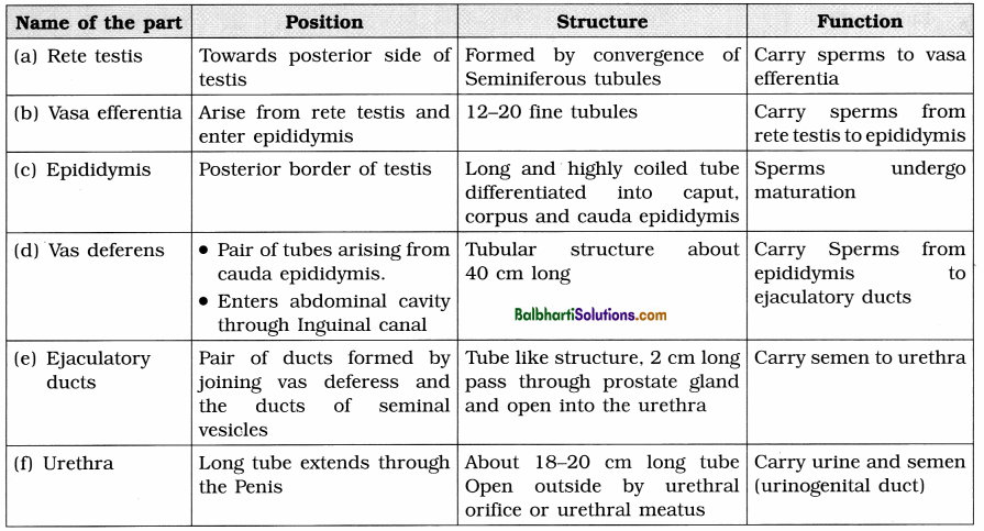
(3) Glands :
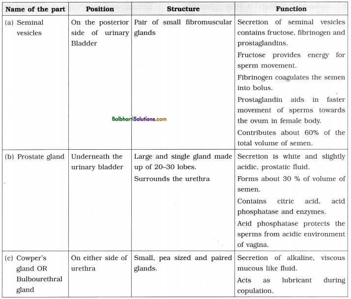
(4) External Ganitalia:
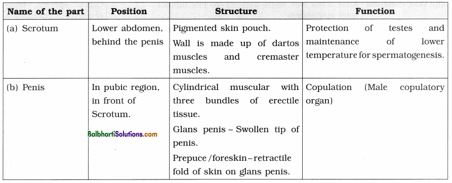
Terms associated with external genitalia of male:
- Inguinal canal : The passage through which testes descend into scrotum.
- Gubernaculum : Fibro-muscular band present in the scrotum.
- Cryptorchidism : Failure of testis to descend into scrotum which may cause sterility.
- Three bundles of erectile tissues in penis :
- Paired Corpora cavernosa
- Median corpus spongiosum.
6. Semen : Single ejaculate of semen is 2.5 to 4.00 ml of viscous, alkaline and milky fluid (pH 7.2 to 7.7) containing 400 million sperms and secretions of epididymis, prostate gland, and Cowper’s glands. It is rich in fructose, Ca++ , bicarbonates and prostaglandins.
7. Histology of testis :
- External coverings of testis seen in L.S. from outer to inner side tunica vaginalis, tunica albuginea and tunica vasculosa.
- Testicular mass divided into 200-300 testicular lobules by tunica albuginea.
- Each lobule has 1-4 seminiferous tubules.
- In between the seminiferous tubules there are interstitial cells of Leydig or Leydig’s cells. They secrete the male hormone androgen or i testosterone.
- Internal lining of cuboidal germinal epithelial cells or spermatogonia inside the seminiferous l tubules.
- Few large pyramidal cells called Sertoli or sub-tentacular cells (nurse cells) Sertoli cells provide nutrition to the developing sperms,
- Various stages of developing sperms such as spermatogonia, primary spermatocyte, secondary spermatocyte, spermatids and sperms are seen in the seminiferous tubules.
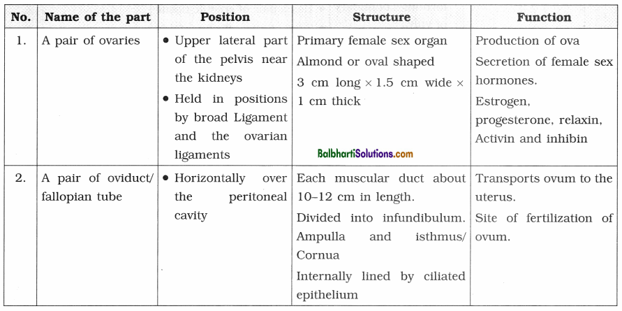
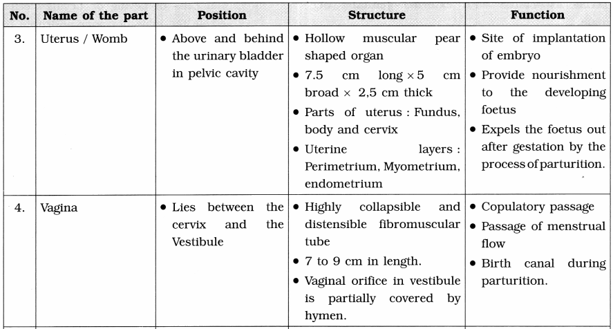
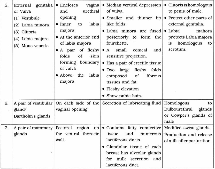
8. Female Reproductive System : The female reproductive system consists of the following parts :
- A pair of ovaries
- A pair of fallopian tubes. (Also called oviducts, but usually this term is used for lower animals)
- Uterus
- Vagina
- External genitalia (vulva)
- A pair of vestibular glands
- A pair of mammary glands
9. Puberty / Sexual maturity in Females :
- Menarche : Onset of menstrual cycle, usually occurs at the age 10-14 years.
- The reproductive system then becomes functional.
- Menopause : End of menstrual cycles in females, usually occurs at the age of 45-50 years.
- Reproductive age of female : The period from menarche to menopause.
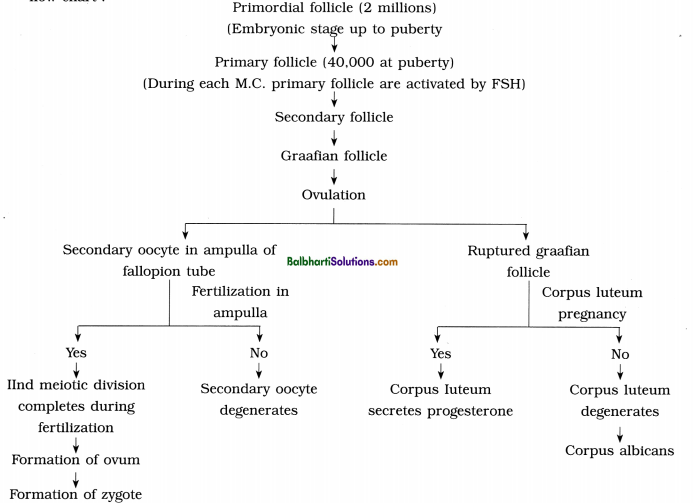
10. Structure and development of the ovary : The process of oogenesis starts in ovary much before the birth of the female baby. The various developing follicles and their fate is shown in the following flow chart :
11. Histology of ovary :
- TWo parts in ovary : Central part is medulla while the peripheral part is called cortex.
- Layer of germinal epithelium covers the cortex.
- Germinal epithelial cells form groups of oogonia in the form of cords called egg tubes of Pfluger which project into the cortex. Each cord ends in egg nest, which is group of oogonial cells. The primordial ovarian follicles develop from these cells.
- Different stages of development of oocyte viz. primordial follicle, primary follicle, secondary follicle, Graafian follicle, rupturing Graafian follicle, corpus luteum, corpus albicans are seen in a cortex of mature ovary.
- Medulla has blood vessels, lymph vessels and theca externa.
- Graafian follicle is a mature ovarian follicle with eccentric secondary oocyte in a fluid filled cavity called antrum surrounded by membrane granulosa, theca interna and theca externa.
- The ovum is surrounded by vitelline membrane, zona pellucida and membrana * granulosa.
- In the antrum, ovum is situated on discus proligerus. Ovum along with discus proligerus ; is called cumulus oophorus. Antrum is filled : with liquor folliculi.
![]()
Menstrual cycle (Ovarian cycle)-
1. Primates show menstrual cycle. Human female shows cycle with 28 days periodicity.
2. It involves a series of cyclic changes in the ovary and uterus.
3. Menstual cycle:
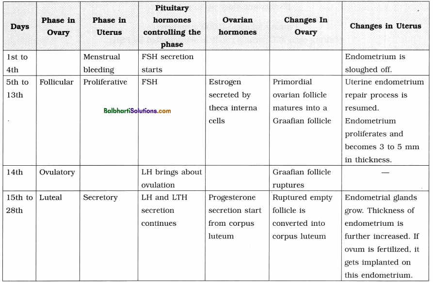
4. If the ovum is fertilized corpus luteum is retained.
5. A once embryo gets implanted in the endometrium, it starts secretion of human chorionic gonadotropin (hCG) which extends the life of corpus luteum.
6. Presence of hCG in maternal blood and urine is an indicator of pregnancy.
7. If ovum is not fertilized, corpus luteum regresses and forms corpus albicans. Then the next menstrual cycle begins.
Gametogenesis-
1. The process of formation of gametes in sexually reproducing animals is called gametogenesis.
2. Sperm is the male gamete and ovum/egg is the female gamete.
3. The gametes are formed from primordial germ cells of gonads.
4. Spermatogenesis : The process by which sperms are formed is called spermatogenesis.
- In the testis, i.e. male gonads there are seminiferous tubules which are lined by germinal epithelium.
- Cells of germinal epithelium undergo spermatogenesis and form sperms.
- In between the germinal cells there are nurse cells or cells of Sertoli which provide nourishment to the developing sperms.
(1) Phases of spermatogenesis : There are three phases of spermatogenesis, viz. multiplication phase, growth phase and maturation phase. Germinal cells are called primordial germ cells.
- Multiplication phase : Repeated mitotic divisions produce large number of spermatogonia which are diploid (2n).
- Growth phase : The spermatogonium accumulates food and grows in size. This is now called primary spermatocyte.
- Maturation phase : The primary spermatocyte undergoes first meiotic division. At the end of this division two haploid secondary spermatocytes are formed. Secondary spermatocyte
undergoes second meiotic division and produce spermatid.
Spermatids are non-motile and non-functional. They undergo metamorphosis in the process of spermiogenesis and mature into motile sperms. Each haploid spermatogonium thus forms four haploid spermatids which later transform into the sperms.
![]()
(2) Changes occurring during spermiogenesis :
- Increase in sperm length
- Distinction of centrioles into proximal and distal one.
- Formation of axial filament from distal centriole
- Formation of spirally coiled mitochondria.
- Formation of acrosome from Golgi complex.
(3) Structure of the sperm (spermatozoa) :
- Microscopic, elongated haploid motile male gamete measuring about 0.055 mm (60 /;) in length.
- Produced by the process of spermatogenesis.
- Their viability remains viable for seventy- two hours, but can fertilize the ovum in first 12 to 14 hours only.
(4) Parts of sperm:
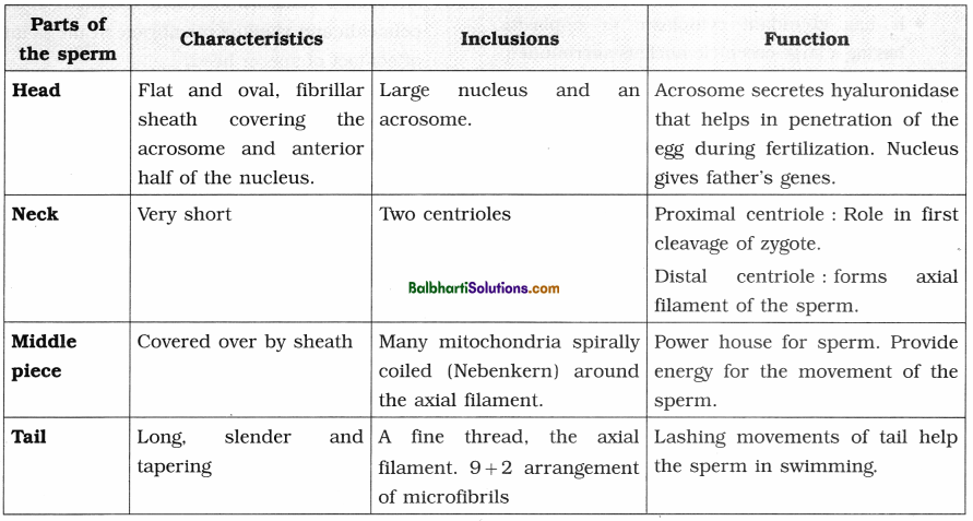
5. Oogenesis : Oogenesis takes place in ovary by which the secondary oocyte is formed.
(1) Phases of oogenesis :
- Phase of multiplication : In the phase of multiplication, large number of oogonia are formed.
- Phase of growth : In the phase of growth, one of the oogonia becomes large. It is now called primary oocyte.
- Phase of maturation : In this phase the rimary oocyte undergoes meiosis-I and forms large haploid secondary oocyte. There is equal nuclear division but unequal cytoplasmic division during the phase of maturation. Secondary oocyte and polar bodies undergo arrested meiotic-I division. Secondary oocyte completes the meiosis-II if and only if it undergoes fertilization.
(2) Structure of ovum, i.e. secondary oocyte : j
- The ovum discharged by the ovary during ovulation is actually a secondary oocyte.
- Rounded, haploid, non-mo tile. Female gamete is largest cell of the body.
- Measures about 0.1 mm or 100 in diameter.
- It is microlecithal, i.e. almost free of yolk.
- It has abundant cytoplasm or ooplasm having a large eccentric nucleus surrounded by vitelline membrane.
- Centriole is absent in the ovum.
- Ovum shows polarity having two poles.
- Animal pole is the side which shows the presence of polar body and nucleus while the opposite side is termed as vegetal pole.
- The ovum is enclosed by two additional coats inner, thin, transparent and noncellular zona pellucida and outer, thick cellular corona radiata.
- Between the vitelline membrane and zona pellucida there is a narrow perivitelline space.
- The zona pellucida is secreted by ovum j itself while corona radiata is formed of radially elongated follicular cells which are glued together by hyaluronic acid.
Fertilization/Syngamy-
1. Fertilization : Process of fusion of the haploid male and female gametes into diploid zygote is called fertilization.
2. Fertilization is internal and in the ampulla of the fallopian tube the gametes meet.
3. The sequence of events of fertilization :
- Capacitation of sperms : Movement of sperms towards the secondary oocyte.
- Release of acrosomal enzymes: Enzymes are secreted by sperm head when it touches secondary oocyte.
- Separation of cells of corona radiata : This happens due to acrosomal enzymes like hyaluronidase and corona penetrating enzymes.
- Compatibility reaction/ Fertilizin-anti fertilizin interaction : Fertilizin of egg binds with anti fertilizin of sperm. The zona pellucida of egg has fertilizin receptor proteins (ZP3, ZP2). Also acrosome ruptures releasing enzyme acrosin / zona lysin.
- Acrosome reaction : Acrosin dissolves zona pellucida and vitelline membrane at the point of contact of sperm head.
- Entry of sperm nucleus in ovum : Nucleus and centriole enter the ovum.
- Formation of fertilization membrane/ Cortical reaction : Vitelline membrane of ovum is converted into a fertilization membrane. This prevents polyspermy.
- Activation of ovum : Completion of meiosis- II of secondary oocyte takes place. When ovum receives centriole from sperm, it completes meiosis-II. This releases second polar body.
- Syngamy/Karyogamy : Fusion of male pronucleus and female pronucleus. Synkaryon is formed after fusion of male and female nucleus.
Formation of diploid zygote conclude the process of fertilization
![]()
4. Significance of fertilization :
- Secondary oocyte can complete meiosis only after fertilization.
- Diploid number of chromosomes is restored in the zygote.
- Genetic characters of two parents can be mixed leading to variation and evolution.
- It determines the sex of young one.
- Only through fertilization centrioles are introduced into the ovum which are otherwise absent in ovum.
Embryonic development-
1. After syngamy the zygote that is formed undergoes divisions to form embryo. These mitotic divisions are called cleavages.
2. When the zygote passes through the fallopian tube, the cleavages start.
3. Till morula stage, zona pellucida layer is retained as it prevents the implantation of the blastocyst at an abnormal site.
4. Cleavage :
- Cleavages are rapid mitotic divisions of zygote to form a hollow spherical multicellular blastula. Cleavage converts the zygote first into a mass of cells called morula.
- Cleavage occurs during its passage through the fallopian tube to the uterus.
- In human beings, cleavage is holoblastic, equal and indeterminate.
- Cleavage divisions are rapid with short interphase.
- There is no time for cells to grow in size.
Thus, cells become progressively smaller. The resulting daughter cells are called blastomeres. - Cleavage shows faster synthesis of DNA.
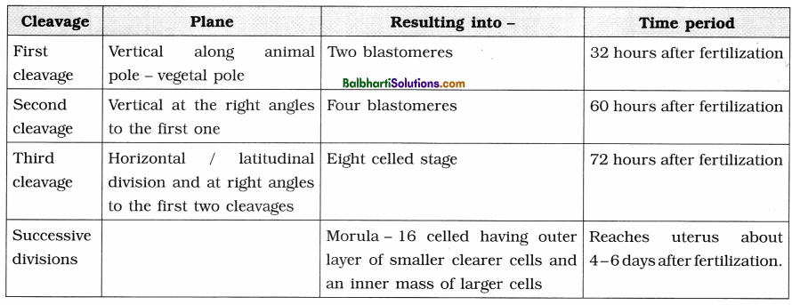
5. Blastulation :
- During blastulation hollow and multicellular blastocyst is formed from 16-32 celled stage of morula.
- Blastocyst now reaches uterus and starts absorbing glycogen rich uterine milk.
- The outer layer of cells forms trophoblast layer whereas inner large cells form inner cell mass or embryoblast.
- Size of blastocyst doubles from 0.15 mm to 0. 30 mm.
- Due to the absorption of glycogen rich uterine milk, the trophoblast cells become flat and a cavity called blastocyst cavity is formed.
- Cells of rauber : These are the cells of trophoblast which are in contact with the embryonal knob.
- Blastocyst becomes fully developed by the end of the 7th day.
6. Implantation :
- Implantation is the process of embedding the blastocyst in the uterine endometrium for further gestation.
- On 7th to 10th day after fertilization, implantation takes place. Embryo is completely buried inside the endometrium.
- Trophoblast divides into two layers, viz. cytotrophoblast and syncytiotrophoblast. With processes of synctiotrophoblast the blastocyst is buried in the endometrium layer of uterus.
7. Gastrulation :
(1) Formation of gastrula from the blastocyst is called gastrulation. It starts at about 8 days after fertilization.
(2) Two important events during gastrulation are :
- Differentiation of blastomeres : Three germinal layers are formed by rearrangement of blastomeres.
- Morphogenetic movements : Movements of cells to reach their destined area of differentiation is called morphogenetic movements.
(3) Gastrulation and implantation of blastocyst takes place simultaneously.
(4) Gastrulation involves the following sequential changes :
- Formation of endoderm
- Formation of embryonic disc
- Formation of amniotic cavity
- Formation of ectoderm
- Formation of mesoderm
- Formation of extra-embryonic coelom
- Formation of chorion and amnion
8. Organogenesis : Process of forming organs after the process of gastrulation is called organogenesis.
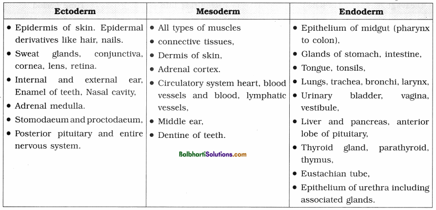
9. Fate of germinal layers : At the end of gastrulation, embryo develops into three germinal layers.
Different tissues and organs are formed from germinal layers. This process is called histogenensis.
Pregnancy / Gestation-
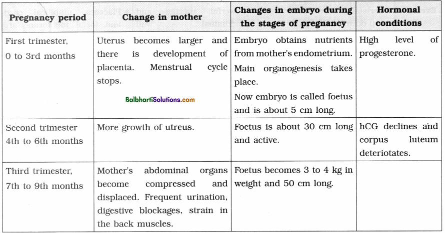
1. Gestation or pregnancy is the condition of developing foetus in the uterus. It may be one or two as in twins.
2. In human beings, gestation period is about 280 days.
Placenta-
- Placenta is a flattened, discoidal organ attached to the wall of the uterus and to the baby’s umbilical cord.
- It facilitates the supply of oxygen and nutrients and also for removal of carbon dioxide and excretory products produced by the foetus.
- Placenta is the only organ, which is formed of tissues from two different individuals the mother and the foetus.
- Foetal placenta is the choronic villi while maternal placenta is the highly vascularized uterine wall. So human placenta is called haemochorial.
- The placenta also acts as an endocrine tissue and produces hormones like hCG, progesterone, estrogen and relaxin.
![]()
Parturition-
- Parturition is the birth process which is accompanied with labour pains.
- It is a neuro-endocrine mechanism which involves rise in estrogen : progesterone ratio and increase in oxytocin receptors in myometrium of uterine wall.
- The fully developed foetus gives signals for the uterine contractions by secreting Adrenocorticotropic hormone (ACTH) from pituitary and corticosteroids from adrenal gland.
- This triggers release of oxytocin from mother’s pituitary gland, which acts on uterine muscles of mother and causes vigorous uterine contractions.
- This leads to expulsion of the baby from the uterus.
- Parturition involves three stages, viz. dilation stage, expulsion stage and after birth or placental stage.


Lactation-
- After parturition the new born is given nourishment through milk. The process of secretion of milk is called lactation in which mammary glands of mother become functional.
- The first milk is called colostrum which is rich in proteins, lactose and mother’s antibodies e.g. IgA.
- Lactation is also neuroendocrine process, in which almost all endocrine glands of mother are involved.
Reproductive Health-
- Total wellbeing of a person’s emotional, behavioural, physical and social aspects involving reproduction is called reproductive health.
- In India, Reproductive and Child Health Care (RCH) programmes are undertaken to improve reproductive health.
- One of the objectives of this programme is to control the population growth of India.
Birth Control-
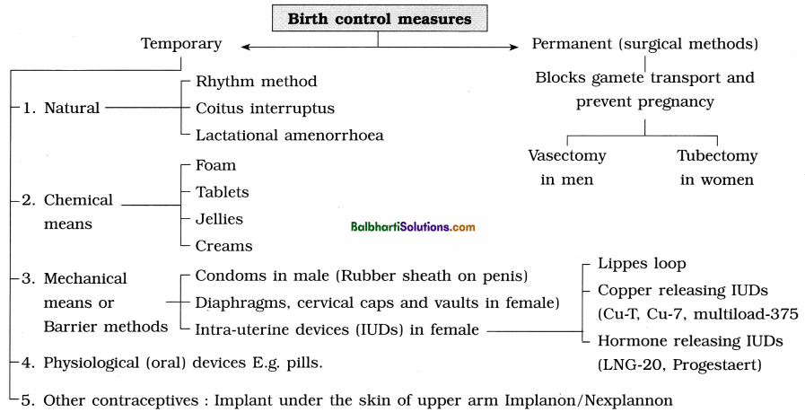
1. For controlling the family size, birth control measures are taken which me called contraceptive measures.
2. Contraceptive methods are of two main types, i.e. temporary and permanent.
Birth control measures:
3. Medical termination of pregnancy (MTP) :
- MTP is induced abortion. This can indirectly control population. MTP is legalized in India since 1971.
- MTP is performed if unwanted pregnancy has to be discontinued or if there are defects in the growing foetus. MTP to abort healthy female embryo is illegal.
- MTP can be safely done only during the first trimester of pregnancy.
4. Amniocentesis :
- Process in which amniotic fluid containing foetal cells is collected using a hollow needle inserted into the uterus under ultrasound guidance.
- This is done for studying the chromosomes to check any possible abnormality in the developing foetus.
- Sex determination by amniocentesis is legally banned in India.
Sexually Transmitted Diseases (STDs)-
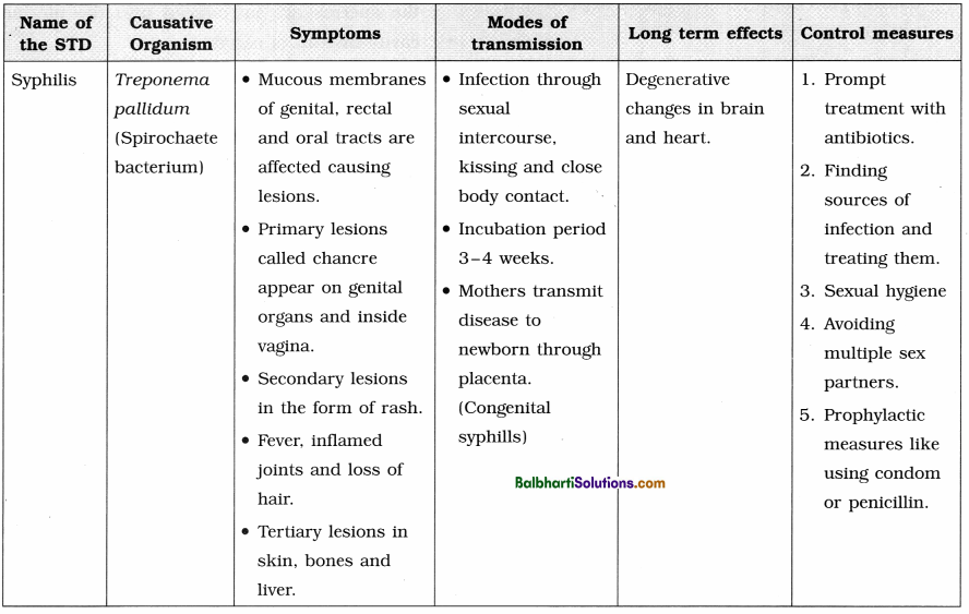
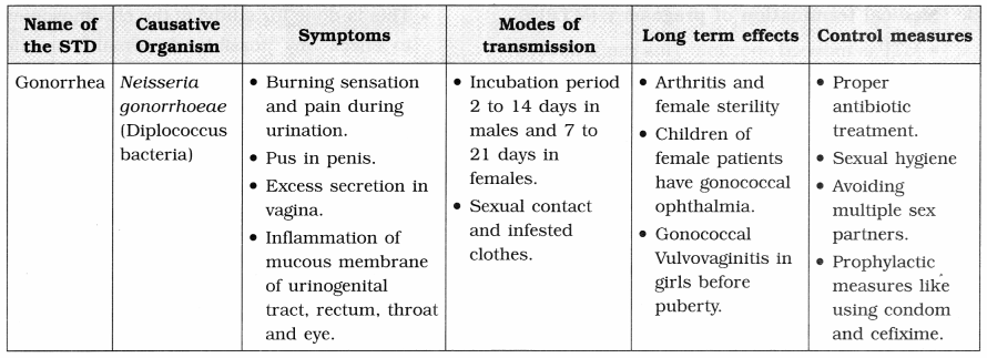
- 1. Disease or infections which are transmitted through sexual intercourse are collectively called Sexually Transmited Diseases (STDs) or Venereal Diseases (VDs) or Reproductive Tract Infections (RTI).
- The major venereal diseases are syphilis and gonorrhoea.
Infertility-
1. Infertility is defined as the inability to conceive naturally after (one year of) regular unprotected intercourse.
2. Today infertile couples have many options to have a child such as fertility drugs, test tube babies, artificial insemination, IUI, surrogate motherhood, etc.
- In Vitro Fertilization (IVF) : When fertilization process is carried out of the body and the embryo is transferred back into the mother’s body, then it is called IVF technique. (Commonly known as test-tube baby.)
- Zygote Intra Fallopian Transfer (ZIFT) : The embryo is transferred in the fallopian tubes by ZIFT (Zygote Intra Fallopian Transfer) technique.
- Gamete Intra Fallopian Transfer (GIFT) : Transferring the ovum collected from the donor into the fallopian tube of another female who can act as a surrogate mother (a female with suitable environment for fertilization and development) is called GIFT.
- ICSI (Intra Cytoplasmic Sperm Injection) : Single sperm cell is injected directly into cytoplasm of an ovum in the laboratory in this technique.
- Artificial insemination (AI) : The collected sperms are artificially introduced into the
cervix of female, for the purpose of achieving a pregnancy through in vivo fertilization. - IUI (Intra Uterine Insemination) : Similar to artificial insemination, but in this technique the sperms are introduced into the uterine cavity instead of cervix.
- Sperm bank / Semen bank : Sperms are collected from donors and stored in a sperm bank or semen bank. These are stored by cryopreservation method and given to needy couples.
- Adoption : A couple or a single parent can legally adopt a child. They also get legal rights, privileges and responsibilities for raising up adopted child.
![]()
Know the Institute :
Cord blood bank, Kolkata
- First Government-run cord blood bank at Kolkata, established in 2001.
- Accredited by AABB (American Association of Blood Bank).
- Work carried out Collection of the cord blood, extracting and cryogenically preserving for its stem cells and other cells of the immune system for future potential medical use
- Stem cells are used to treat diseases, e.g. leukaemia, certain cancers, sickle-cell anaemia and some metabolic disorders.