By going through these Maharashtra State Board 12th Science Biology Notes Chapter 8 Respiration and Circulation students can recall all the concepts quickly.
Maharashtra State Board 12th Biology Notes Chapter 8 Respiration and Circulation
Respiration-
- Respiration is a biochemical process of oxidation of organic compounds in an orderly manner for the liberation of chemical energy in the form of ATP
- C6H12O6 + 6O2 → 6CO2 + 6H2O + 38ATP
![]()
Organs of respiratory exchange-
1. Respiratory surface should possess the following features for efficient gaseous exchange.
- A large surface area.
- Thin, highly vascular and permeable to allow exchange of gases.
- Moist surfaces.
2. A terrestrial plant has stomata on leaves and young stems and lenticels on the stem surface for exchange of gases.
3. In animals, depending upon the complexity of organization and the surrounding medium, respiratory organs have become specialized and are usually associated with a transport system.
4. Respiratory organs In different organisms:
| Organism | Respiratory surface or organ |
| I. Aquatic organisms | |
| (1) Protists, sponges, coelenterates | Plasma membrane |
| (2) Planaria, Annelids, Amphibians | Plasma membrane, general body surface and moist skin |
| (3) Limulus (Arthropod) | Book gills |
| (4) Amphibian tadpoles, salamander and newt | External gills |
| (5) Fish | Internal gills |
| Organism | Respiratory surface or organ |
| II. Terrestrial organisms | |
| (1) Insects (2) Arachnids (Spider and Scorpion) (3) Reptiles, birds and mammals | Tracheal tubes and spiracles Book lungs Lungs |
| III. Underwater organism | |
| Turtle | * Cloaca |
| Learn this as well : Only at the time of diving or when underwater, turtles perform cloacal respiration. There are a pair of accessory air bladders connected to the cloaca which can absorb oxygen from the water. | |
![]()
Human respiratory system-
Human respiratory system consists of nostrils, nasal chambers, pharynx, larynx, trachea, bronchi, bronchioles, lungs, aided by diaphragm and intercostal muscles.
1. Nostrils and nasal chambers :
(1) The nostrils are external openings of the nose. Oxygen rich air is taken into the body through the nostrils or external nares. Carbon dioxide and water vapour are released out of the body through the nostrils.
(2) The internal nares open into the pharynx. The space between the external and internal nares is known as nasal chamber. The nasal chamber is lined internally by mucous membrane and ciliated epithelium.
(3) The nasal chamber is divided into right and left parts by a cartilage called mesethmoid. Every nasal chamber is further divided into three regions, viz. vestibule, respiratory part and sensory part.
- Vestibule : The anteriormost part of the nasal chamber is vestibule. Hair present in this chamber prevent the dust particles from going inside.
- Respiratory part : It is the part which is richly supplied by capillaries. Air is made warm and moist in this region.
- Sensory part : The sensory epithelium lines this region. It is concerned with the detection of smell.
2. Pharynx :
- The pharynx is a short, vertical tube about 12 cm in length. The respiratory and food passages cross each other in the pharynx.
- The upper part of the pharynx is known as naso-pharynx. It conducts the air.
- The lower part is called laryngo-pharynx or oro-pharynx. It conducts food to the oesophagus.
- The tonsils are present in the pharynx. They are made of lymphatic tissue. They kill the bacteria that are trapped in mucus.
3. Larynx :
- The larynx produces sound. In males, it increases in size at puberty. This is termed as Adam’s apple.
- From the pharynx air enters the larynx. The opening through which it enters is called glottis.
- The glottis has a guarding flap called epiglottis.
- The epiglottis prevents the entry of food particles into the trachea.
- The vocal cords are seen along the side of the glottis. They are made of elastic tissue. They produce sound.
- Passage of air between the vocal cords and modulations created by tongue, teeth, lips and nasal cavity produce voice.
4. Trachea :
- The trachea or windpipe is about 10-12 cm long and 2.5 cm wide.
- It is situated in front of the oesophagus and runs downwards in the thorax.
- Fibrous muscular tissue supported by ‘C’ shaped cartilages form the walls of the trachea.
- 16 to 20 cartilage rings are present in the trachea.
- The trachea is lined internally by ciliated epithelium and mucous glands.
- Mucous and ciliary action remove the dust particles and push them upwards to the larynx. These particles are then gulped and taken into the oesophagus. Instant coughing can remove foreign particles that enter the trachea.
5. Bronchi and bronchioles :
- The trachea divides into two bronchi (singular- bronchus) at its distal end behind the sternum.
- The bronchus has complete ring of cartilage for support.
- The bronchi enter the lungs on either side.
- After entering the lungs each bronchus divides into secondary and tertiary bronchi. The tertiary bronchi divide further to form bronchioles.
- The bronchioles are minute and are without the cartilage rings in their walls.
- Each bronchiole ends into a bunch of alveoli which appear like a bunch of grapes. Each alveolus is balloon shaped.
- Many alveoli make the lung spongy and elastic.
6. Lungs :
- The lungs are principal respiratory organs located in the thoracic cavity.
- They are pinkish, soft, hollow, paired, elastic and distensible organs.
- The lung is enclosed in a pleural sac.
- The pleural sac has two membranes – an outer parietal and an inner visceral which enclose the pleural cavity.
- The pleural fluid which is present in the pleural cavity lubricates and prevents friction when pleural membranes slide on each other.
- The lungs are richly supplied with blood capillaries and hence are highly vascular organs.
- The left lung has two lobes while the right lung has three lobes.
- Each lobe has many bronchioles and alveolar sacs.
- The alveolar sacs are spherical and thin walled and contain about 20 alveoli.
- The alveoli are covered by a network of capillaries from pulmonary artery and pulmonary vein.
- Each alveolus has thin and elastic wall. It is about 0.1 mm in diameter.
- The alveolar wall is 0.0001 mm thick and is made of simple, non-ciliated, squamous epithelium. It has collagen and elastin fibres.
- Every lung has about 700 million alveoli which increase the surface area for the exchange of gases.
- The outermost covering of the lungs which is known as visceral pleura is made of smooth muscle fibres.
- The lobule in the lung consists of alveolar ducts, alveolar sacs and alveoli. In alveoli gaseous exchange takes place.
![]()
Mechanism of respiration –
1. The process of respiration includes breathing, external respiration, internal respiration and cellular respiration.
(A) Breathing :
- Breathing is the process by which the air comes in and goes out of the lungs.
- The rate of gaseous exchange is speeded up by breathing.
- Breathing is a part of respiration and the terms breathing and respiration are not synonymous.
- Inspiration and expiration together make breathing.
(i) Inspiration :
- Inspiration is an active process brought about by ribs, intercostal muscles, sternum and diaphragm.
- The intercostal muscles contract and pull the ribs outwards. This increases the space in the thoracic cavity. The lower part of sternum is simultaneously raised. The diaphragm contracts and flattens. This
causes further increase in the volume of : thoracic cavity. - Pressure in the lungs decreases and the volume | increases due to expansion of the lungs.
- Due to pressure difference the atmospheric * air rushes into the lungs through respiratory * passage as a result of which air is inspired * in.
(ii) Expiration :
- Expiration is the passive process.
- During expiration the intercostal muscles j relax and the ribs are pulled inwards.
- The diaphragm is relaxed and becomes ; dome-shaped.
- The volume of the thoracic cavity is reduced. :
- The pressure on the lungs is increased as a j result of which they get compressed.
- Air is thus expelled out of lungs through the j nares.
(iii) Respiratory cycle :
- Respiratory cycle is alternate inspiration and expiration process.
- In adult man there are 16 to 20 respiratory cycles per minute.
- The medulla oblongata in the brain controls the respiration.
(B) External respiration/exchange of gases at the alveolar level :
- Exchange of gases between the alveolar .air and the blood takes place through thin squamous epithelial layer of alveolus and similar layer of the capillary wall.
- Respiratory gases will always diffuse from an area of higher partial pressure to an area of lower partied pressure in these two regions.
- Due to difference in partial pressure, carbon dioxide diffuses from the capillaries into the alveolus whereas oxygen will diffuse from alveoli to the capillaries.
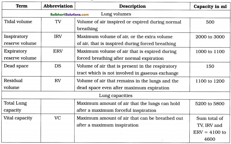
- Table : Pulmonary volumes and capacities :
(C) Internal respiration :
- Transport of O2 : Only 3% of total oxygen is carried in a dissolved state by plasma while 97% of oxygen is carried in the form of oxyhaemoglobin from lungs to tissues.
- Oxygen dissociation curve : A sigmoid curve which shows oxygen-haemoglobin dissociation and the relationship between oxyhaemoglobin saturation and oxygen tension.
- Bohr effect : The shift of oxyhaemoglobin dissociation curve due to change in partial pressure of C02 in blood is called Bohr effect.
- Haldane effect : The effect caused by increase in hydrogen ions which results in decrease of pH of blood is called Haldane effect.
Transport of CO2 :
- 7% of COa is transported in the form of carbonic acid by plasma.
- 70% of COa is transported from tissues to lungs in the form of sodium bicarbonate and potassium.
- Remaining 23% of COa is carried in the form of carbaminohaemoglobin.
- Hamburger’s phenomena or chloride shift : Movement of chloride ions to maintain the ionic balance between the RBCs and the plasma is called chloride shift.
(D) Cellular respiration : In this last step food is oxidized in the cell and ATP is produced and used to carry out vital processes.
2. Carbon monoxide poisoning :
- Haemoglobin has affinity for oxygen. But for carbon monoxide it has about 250 times more affinity than that of oxygen.
- With carbon monoxide it forms a stable compound called carboxyhaemoglobin.
- Due to such combination, the oxygen is not transported to the tissues. The tissues thus suffer from oxygen starvation. This leads to asphyxiation and in extreme cases death.
- Treatment of carbon monoxide poisoning is given by administering oxygen-carbon dioxide mixture to make high PO2 level to dissociate the carbon monoxide from haemoglobin.
- Carbon monoxide poisoning occurs in closed rooms with open stoves, gas burners, automobile engines or any incomplete combustion.
![]()
Regulation of breathing-
- Normal breathing is an involuntary process controlled by inspiratory centres and expiratory centres in medulla, pneumotaxic centre in pons and apneustic centre located in medulla.
- The Hering-Breuer reflex controls the rate and depth of breathing and also prevents over inflation of lungs.
- Cerebral cortex has voluntary centres which prevent water or irritating gases from entering the lungs.
Modified respiratory movements-‘
Modified respiratory movements are used to express emotions and to clear air passages. They may be reflexes or voluntarily initiated movements such as yawning.
Common disorders of respiratory system-
1. Respiratory disorders:
| Respiratory disorders | Cause and symptoms |
| (1) Emphysema | Cause : Cigarette smoking and air pollution Symptoms : Over inflation of the alveoli, rupture of alveolar wall. |
| (2) Bronchitis | Cause : Certain bacterial or viral infection, also caused by smoking and air pollution. Symptoms : Inflammation of bronchi, regular coughing with greenish-yellow sputum. |
| (3) Sinusitis | Cause : A viral infection or common cold Symptoms : Inflammation or swelling of the tissue lining the sinuses. |
| (4) Laryngitis | Cause : Certain viruses, bacteria Symptoms : Hoarseness, cough, difficulty in swallowing, inflammation of larynx and vocal cords. |
| (5) Pneumonia | Cause : Bacteria, viruses, mycoplasma Symptoms : Filling of air spaces of alveoli with fluid containing dead WBCs, chest pain, shortness of breath, blood in mucous. |
| (6) Asthma | Cause : Allergy to foreign substances like pollen, dust, certain food, food additives, animal dander, etc. Symptoms : Narrowing and inflammation of bronchi, bronchospasm, periodic wheezing, difficulty in breathing. |
| (7) Occupational respiratory disorders silicosis, asbestosis | Cause : Long term exposure to silica and asbestos dust in the mining industry. Symptoms : Irritation, fibrosis causing inflammation. |
2. Treatment of respiratory disorders is by taking suitable antibiotics, inhalants, vaporizers and cough medicines. Also quitting smoking, using preventive masks and staying away from polluted air is too remedy against these disorders.
3. Artificial ventilation : Method of induced breathing in a person who is unable to breathe is given artificial ventilation.
4. Ventilator : A machine supporting breathing when normal breathing fails.
![]()
Transportation in living organisms-
Circulation in animals-
1. Transportation by diffusion and active transport is suitable in extremely small organisms.
2. Intracellular transport by cyclosis is shown by almost all living organisms e.g. Paramoecium, Amoeba, root hair cells of many plants and WBCs in animals.
3. Extracellular transport : In this transport water or body fluid is circulated through body cavities as in sponges and coelenterates or moved around the viscera by contraction of body wall and muscles as in roundworms or parenchymal circulation, viz. flatworms.
4. Blood vascular system in higher animals from Annelida to chordate contains
- blood as a circulating fluid,
- heart as a pumping organ and
- the blood vessels through which blood circulates.
5. Types of blood vascular system :
(1) Open circulation :
- In this type, blood finally conies out of the blood vessels and is circulated through the body cavities (haemocoel).
- Blood flows at low pressure and there is direct exchange of materials between blood and cells or tissues of the body.
- Respiratory pigment is usually absent. When present, it is dissolved in plasma of the blood, e.g. Arthropods and Molluscs.
(2) Closed circulation :
- In this type of circulation, blood is circulated all over the body through the network of blood vessels.
- Blood does not come in direct contact with cells and body tissues and the exchange of materials between the blood and cell takes place through an intermediate fluid called lymph.
- Blood flows through blood vessels at high pressure and can be regulated. Respiratory pigment like haemoglobin is present for transportation of respiratory gases, e.g. All vertebrates, higher molluscs and annelids.
- Closed circulation can be of two main types : single circulation and double circulation.
(a) Single circulation : In fishes heart shows single circulation as blood passes only once through heart during one cardiac cycle.
(b) Double circulation :
- Human heart shows double circulation as blood passes twice through the heart during one cardiac cycle. The blood follows two routes, viz. pulmonary and systemic.
- Pulmonary circulation is the circulation between the heart and the lungs. The course of blood during pulmonary circulation is from the right ventricle (by pulmonary trunk) to the left atrium (by two pairs of pulmonary veins) of heart through lungs.
- Systemic circulation is the circulation between the heart and the body organs (except lungs). The course of blood during systemic circulation is from left ventricle (by systemic aorta) to all body organs and from the body back to right atrium (by vena cavae).
Learn This As Well:
Coronary circulation is circulation to the cardiac muscles of the heart. Coronary arteries supply oxygenated blood whereas coronary veins join to form coronary sinus and collect deoxygenated blood. This sinus opens into the right atrium.
Circulatory System in Human-
1. Circulatory system in human is made up of blood vascular system and lymphatic system.
2. Blood vascular system consists of blood, heart and blood vessels.
3. Blood composition and Coagulation :
- Study of blood is called haematology.
- The bright red, slightly alkaline main circulating, fluid in the human body is blood.
- Blood is a fluid connective tissue derived from mesoderm. It has pH about 7.4.
- There are about 5 litres of blood in the body which is about 8% of the total body weight.
- Composition of blood : There are two main components of blood, viz., plasma (55%) and blood corpuscles (45%).
(i) Plasma : Plasma is a straw coloured fluid part of blood, slightly alkaline, viscous fluid consisting of 90 – 92% water and 8 – 10% of solutes.
- Solutes are 7% proteins (serum albumin, serum globulin, heparin, fibrinogen and prothrombin).
- Other solutes are nutrients (glucose, amino acids, fatty acids and glycerol).
- Nitrogenous wastes such as urea, uric acid, ammonia and creatinine.
- Gases like oxygen, carbon dioxide and nitrogen.
- Regulatory substances like enzymes and hormones.
- Inorganic substances like bicarbonates, chlorides, phosphates and sulphates of sodium, potassium, calcium, magnesium, etc.
(ii) Blood corpuscles : Blood corpuscles are of three types, viz. erythrocytes (RBCs), leucocytes (WBCs) and thrombocytes (platelets).
![]()
Red blood corpuscles/erythrocytes-
- Circular, biconcave, enucleated cells of about 7 /im in diameter and 2.5 /mi in thickness.
- RBC count is about 5.1 to 5.8 million RBCs/ cu mm in male and 4.3 to 5.2 million/cu mm in female. The average life span of RBC is about 120 days.
- Erythropoiesis is formation of RBCs. It occurs in liver and spleen in foetus and in red bone marrow in adults.
- The old RBCs are destroyed in liver and spleen.
- Polycythemia is increase while erythrocytopenia is decrease in number of RBCs.
- RBCs contain respiratory pigment called haemoglobin which helps in transport of oxygen and carbon dioxide.
- The normal haemoglobin content in adult male is 14-17 gm/100 ml of blood and 13-15 gm/100 ml of blood in adult female.
- Less amount of haemoglobin leads to anaemia.
- RBCs transport oxygen from lungs to tissues and carbon dioxide from tissues to lungs. They maintain blood pH as haemoglobin acts as a buffer. They also maintain the viscosity of the blood.
- RBCs also contains an enzyme, carbonic anhydrase.
- The haematocrit is the ratio of the volume of RBCs to total blood volume of blood. Its value is different in men and women.
White blood corpuscles/Leucocytes-
- Leucocytes are colourless, nucleated, amoeboid and phagocytic cells.
- They show diapedesis, i.e. squeezing out of blood capillaries by amoeboid movement.
- The size is about 8 to 15 fxm.
- Total WBC count is 5000 to 11000 WBCs/ cu mm of blood.
- The average life span of WBCs is about 3 to 4 days.
- Leucopoiesis or formation of WBCs. It occurs in red bone marrow, spleen, lymph nodes, tonsils, thymus and Payer’s patches.
- Leucocytosis is increase while leucopenia is decrease in the number of WBCs.
- Leukaemia or blood cancer is a pathological increase in number of WBCs.
- The dead WBCs are destroyed by phagocytosis in blood, liver and lymph nodes.
- Leucocytes are of two types, viz., granulocytes and agranulocytes.
- Granulocytes are of three types, viz. neutrophils, eosinophils and basophils.
- Agranulocytes are of two types, viz. monocytes and lymphocytes.
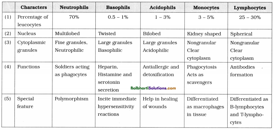
- Characteristics of different types of leucocytes
Thrombocytes/Platelets-
- Thrombocytes are smallest, non-nucleated, round and biconvex. They are of about 2.5 to 5 m in diameter. Their count is about 2.5 – 4.5 lakhs/cu mm.
- Their life span is about 5 to 10 days.
- Megakaryocytes of bone marrow form thrombocytes.
- Thrombopoiesis is the formation of platelets.
- Thrombocytosis is the increase while thrombocytopenia is the decrease in platelet count.
- Thrombocytes help in coagulation of blood by releasing thromboplastin.
- Blood clotting/coagulation of blood : Active anticoagulants like heparin and antithrombin are present in the intact blood vessels. But upon the rupture of a blood vessel, bleeding starts. The fluid blood is converted into semisolid jelly by the process of blood coagulation or clotting.”
The clotting of blood is a complicated process in which many factors (12 clotting factors) present in plasma and tissues are involved.
The event that take place during blood clotting are as follows :
- Release of thromboplastin from extrinsic source in tissue and intrinsic source in plasma at injured site through a step-wise (cascade process) process.
- Formation of enzyme prothrombinase in the blood.
- Conversion of prothrombin into thrombin by prothrombinase.
- Conversion of fibrinogen into fibrin by thrombin.
- Formation of mesh by the fibrin fibres forming the clot.
- The normal clotting time is 2 to 8 minutes.
![]()
Heart-
1. Heart is a hollow, muscular, conical organ about the size of one’s fist with broad base and narrow apex tilted towards the left.
2. It is mesodermal in origin.
3. It is situated in middle of the thoracic cavity in a space called mediastinum, between the two lungs.
4. The heart is 12 cm in length, 9 cm in breadth and 250 to 300 grams in weight.
5. Pericardium : Double layered membrane, these layers are as follows :
- Fibrous pericardium : Outer, tough layer of inelastic fibrous connective tissue.
- Serous pericardium : This inner pericardium has two layers, outer parietal layer and inner visceral layer.
- Parietal layer forms the inner lining of fibrous pericardium.
- Visceral layer or epicardium is next to heart on the outer side.
- Pericardial fluid is present between the parietal and visceral layers of serous pericardium.
6. Heart wall :
- The heart wall has three layers, viz. outer epicardium, middle myocardium and inner endocardium.
- Epicardium has single layer of flat epithelial cells called mesothelium.
- Myocardium has cardiac muscle fibres responsible for movements of the heart.
- Endocardium has single layer of flat epithelial cells called endothelium.
7. External structure of heart :
- Human heart consists of two superior, small, thin walled receiving chambers called atria or auricles and two inferior, large, thick walled, distributing chambers called ventricles.
- Atrio-ventricular groove or coronary sulcus, a transverse groove which is present between the atria and the ventricles is seen externally.
- The interventricular sulcus is present between the right and left ventricles. Coronary arteries and coronary veins are present in the sulci. The coronary veins join to form coronary sinus which opens into the right atrium.
- The right atrium receives deoxygenated blood from all over the body through superior vena cava and inferior vena cava.
- Left atrium receives oxygenated blood from lungs through two pairs of pulmonary veins.
- From the right ventricle deoxygenated blood is sent to lungs through pulmonary trunk.
- From the left ventricle oxygenated blood is sent to entire body by systemic aorta.
- Ligamentum arteriosum connects the pulmonary trunk and systemic aorta. It represents ductus arteriosus of foetus.
8. Internal structure of heart :
- There are four chambers in the heart, viz., two atria and two ventricles which can be demarcated internally.
- Atria are thin walled upper receiving chambers separated from each other by interatrial septum.
- The right atrium receives deoxygenated blood from all over the body through superior vena cava, inferior vena cava and from the heart through coronary sinus.
- The opening of inferior vena cava is guarded by Eustachian valve while the opening of coronary sinus is guarded by Thebesian valve.
- The fossa ovalis is oval depression that is present on the right side of interatrial septum. It is the remnant of foramen ovale, an oval opening in the interatrial septum of the foetus.
- The left atrium receives oxygenated blood from the lungs through four openings of pulmonary veins.
- Right and left atria open into the right and left ventricles respectively through atrioventricular apertures. These are respectively guarded by tricuspid and bicuspid valves made up of connective tissue.
- The right atrioventricular valve has three flaps hence called tricuspid valve while left atrioventricular valve has two flaps hence called bicuspid valve or mitral valve.
- These valves are attached to papillary muscles of ventricles by chordae tendinae. The valves are prevented from turning back into the atria during the contraction of ventricles due to chordae tendinae.
- Ventricles are two thick walled lower, distributing chambers separated from each other by interventricular septum.
- Left ventricle has thick wall. The inner surface of the ventricle is thrown into a series of irregular muscular ridges called columnae carnae or trabeculae carnae.
- Pulmonary trunk or aorta arises from the right ventricle carrying deoxygenated blood to lungs for oxygenation. Systemic aorta arises from the left ventricle carrying oxygenated blood to all parts of the body.
- Pulmonary aorta and systemic aorta have three semilunar valves at the base which prevent the backward flow of blood during ventricular diastole.
9. Pumping action of heart : Heartbeat is the rhythmic contraction, i.e. rhythmic contraction (systole) and relaxation (diastole) of the heart. The rate of heartbeat is about 72 times per minute during which it pumps out about 5 litres of blood which equals cardiac output.
10. Conducting system of heart :
- The heartbeat in human beings originates in modified cardiac muscles called sinoatrial node (S.A. node). Therefore, the heart is said to be myogenic.
- The conducting system of heart consists of sinoatrial node (SAN), atrioventricular node (AVN), Bundle of His and Purkinje fibres.
- The heart shows auto-rhythmicity as the impulse for its rhythmic movement during beating is developed inside the heart.
- The autorhythmic fibres are developed during embryonic life. They act as pacemaker by setting the rhythm for the heart. They also form conducting system for conducting impulses throughout heart muscles.
- The impulse travels in the heart in the following manner : Sinoatrial node (Pacemaker) → Internodal pathway → Atrioventricular node → Bundle of His → Right and left bundle branches → Purkinje fibres.
![]()
Working mechanism of human heart-
1. Cardiac cycle : One atrial systole (0.1 second), one ventricular systole (0.3 second), followed by a joint diastole (0.4 second) is called a cardiac cycle. One cardiac cycle takes place in about 0.8 second and is also called a heartbeat.
(1) Atrial systole : During atrial systole, the deoxygenated blood from the right atrium enters the right ventricle through atrioventricular aperture whereas the oxygenated blood from left atrium enters the left ventricle through atrioventricular aperture. In normal conditions atrial systole lasts for 0.1 second and atrial diastole lasts for 0. 7 second.
(2) Ventricular systole : During ventricular systole, the deoxygenated blood from the right ventricle enters the pulmonary trunk and the oxygenated blood from the left ventricle enters the aorta. The backflow of blood into atria is prevented by the closure of cuspid valves of both atrioventricular apertures (lubb sound is produced) Ventricular systole lasts for 0.3 second and ventricular diastole lasts for 0.5 second.
(3) Joint diastole or complete diastole : Both atria and ventricles undergo relaxation. During ventricular diastole the backflow of blood from pulmonary trunk and systemic aorta into respective ventricles is prevented by closure of semilunar valves (dub sound is produced). The joint diastole lasts for 0.4 second.
2. Regulation of cardiac activity :
- Cardiovascular centre present in the medulla oblongata of brain regulates the working of the heart.
- Sympathetic nerves secrete adrenaline, which increases the rate of the heart.
- Parasympathetic nerves secrete acetylcholine, which decreases the rate of the heart.
- Conditions like hypoxia, acidosis, alkalosis decrease cardiac activity whereas hormones like epinephrine and nor epinephrine increase cardiac activity (chemical control).
- Elevated level of K+ and Na+ decreases cardiac activity.
Blood vessels-
1. Blood vessels are of three types, viz. arteries, veins and capillaries.
- Arteries : Blood vessels carrying blood away from the heart are called arteries. Arteries form arterioles which in turn divide and re¬divide to form capillaries.
- Veins : Blood vessels carrying blood to the heart are called veins. They have broad lumen and show low blood pressure.
- Capillaries : Capillaries are thinnest of blood vessels and formed by division and redivision of arteriole. Capillaries unite to form venules. Venules join to form veins.
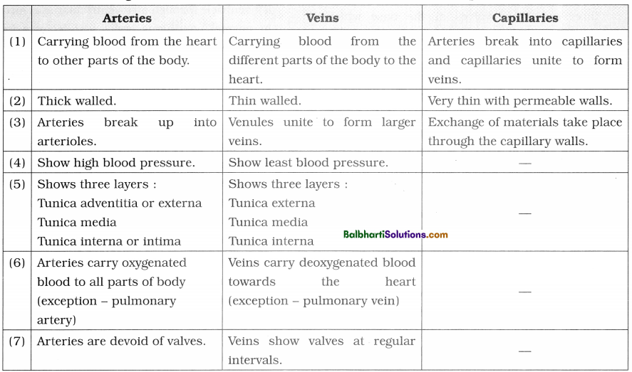
2. A chart showing the differences between arteries, veins and blood capillaries :
Angiology is the study of blood vessels.
3. Heartbeat, pulse and cardiac output :
- Heartbeat is the rhythmic contraction and relaxation of the heart.
- One systole and one diastole make one heartbeat.
- Heart rate is number of beats per minute (72 times per minute).
- Stroke volume is amount of blood pumped out of the ventricles each time (About 70 ml of blood).
- Cardiac output is amount of blood pumped out of the ventricles per minute,
i. e 72 x 70 ml = 5040 or about 5 litres of blood per minute. - Tachycardia is faster heart rate (Over 100 beats per minute).
- Bradycardia is slower heart rate (Over 60 beats per minute).
- Pulse is a pressure wave travelling through the arteries after each ventricular systole.
- Pulse in the radial artery at the wrist is commonly measured.
- The pulse rate per minute indicates the heart rate. It is same as that of heart rate (72 per minute).
- Pulse rate changes with age, sex, posture, exercise and emotional state.
Blood pressure (B.P.)-
1. Blood pressure : Arterial blood pressure is the lateral pressure or force exerted by flowing blood on the wall of arteries.
2. Sphygmomanometer is the instrument used for measuring the blood pressure.
3. The units of blood pressure are mm Hg millimetres of mercury).
4. Blood pressure is of two types-systolic blood pressure and diastolic blood pressure.
- Systolic blood pressure : It is the maximum pressure of blood during ventricular systole. Normal systolic pressure is 120 mm Hg.
- Diastolic blood pressure : It is the minimum pressure of blood during ventricular diastole. Normal diastolic pressure is 80 mm Hg.
- The normal blood pressure is 120/80 mm Hg.
- Pulse pressure is the difference between systolic and diastolic pressure. Normal pulse pressure is 40 mm Hg.
5. Factors affecting arterial blood pressure :
- Cardiac output
- Peripheral resistance
- Blood volume
- Length and diameter of blood vessels
- Viscosity of blood
- Age
- Gender
- Venous return
- Sleep, emotions
- Exercise, anxiety
![]()
6. Hypertension :
- Hypertension means higher values of blood pressure (More than 140/90 mm Hg blood pressure values).
- Excessive high blood pressure of about 230/120 mm Hg may cause rupturing of blood vessels of eye (causing blindness), kidney (nephritis) and brain (stroke or paralysis).
- Factors such as arteriosclerosis, atherosclerosis, obesity, physical , or emotional stress, alcoholism, smoking, cholesterol rich diet, increased secretion of renin, epinephrine or aldosterone, etc. can cause blood pressure.
7. Coronary artery disease (CAD) :
- Atherosclerosis (narrowing of coronary arteries) can cause coronary artery disease.
- In CAD the heart muscle is damaged because of an inadequate amount of blood due to obstruction of its blood supply.
- Depending on the degree of obstruction symptoms may be mild chest pain (angina pectoris) or heart attack (myocardial infarction).
8. Atherosclerosis : Deposition of fatty substances in the lining of arteries, resulting in the formation of an atherosclerotic plaque. These depositions decrease the size of the arterial lumen.
9. Angina pectoris : Angina pectoris is the pain in the chest due to reduction in blood supply to cardiac muscle caused by narrowed and hardened coronary arteries.
10. Angiography : Angiography is X-ray imaging of the cardiac blood vessels to locate the position of blockages. Remedied procedures like angioplasty or bypass surgery are carried out depending upon the degree of blockage.
11. Heart Transplant : Heart transplant is replacement of severely damaged heart by normal heart from brain-dead or recently dead donor. This procedure is necessary in patients with end-stage heart failure and severe coronary arterial disease.
12. Silent heart attack : Heart attack that lacks the general symptoms of classic heart attack like extreme chest pain, hypertension, j shortness of breath, sweating and dizziness is known as silent heart attack or silent myocardial infarction. Men are more affected J by silent heart attack than women.
Electrocardiogram-
- Electrocardiogram or ECG is graphic record : of electrical variations produced by the heart during one heartbeat or cardiac cycle.
- Electrocardiogram or ECG machine is the instrument used to record action potentials generated by heart muscles.
- Einthoven in 1903 discovered this technique, hence he is known as the “Father of Electrocardiography”.
- A normal ECG consists of different types of waves such as P-wave, QR S-complex wave and T-wave.
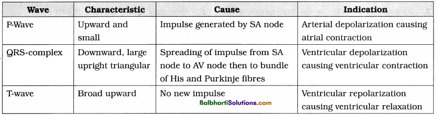
- Waves of ECG and their significance :
- Abnormal functioning of heart such as in coronary artery diseases, heart block, angina pectoris, tachycardia, ischemic heart disease, myocardial infarction, cardiac arrest, etc. can be diagnosed by ECG.
Lymphatic system-
1. Lymph, lymphatic capillaries, lymphatic vessels and lymph nodes together constitute lymphatic system.
- Lymph is the tissue fluid that bathes the cells and is collected in lymphatic capillaries. Lymph is a fluid connective tissue just like blood but is without RBCs, platelets and some plasma proteins. It contains carbon dioxide and metabolic wastes.
- Lymphatic capillaries are thin walled vessels interwoven with the blood capillaries, present in all the tissue spaces. They are not connected with blood capillaries and are blind at one end. Lymph capillaries are wider than blood capillaries and are lined by endothelium of thin and flat cells.
- Lymphatic vessels are formed by the union of lymphatic capillaries. These are thin walled having numerous valves to prevent backflow. Thoracic or left lymphatic duct and right lymphatic duct are the main lymphatic vessels in the body.
![]()
2. Functions of lymphatic system :
- Draining off the excess tissue fluid from the extracellular spaces back into the blood.
- Transport of carbon dioxide and metabolic wastes from the tissue fluid. Transport of lymphocytes and antibodies from the lymphatic nodes to the blood.
- Transport of absorbed fats from the intestine to the blood.
- Destruction of invading microorganisms and foreign particles in the lymph nodes.