By going through these Maharashtra State Board 12th Science Biology Notes Chapter 9 Control and Co-ordination students can recall all the concepts quickly.
Maharashtra State Board 12th Biology Notes Chapter 9 Control and Co-ordination
Introduction-
- Plants show control and coordination by sending chemical signals and bringing about various types of movements.
- Animals control and coordinate the body’s activities by electrical and chemical signals.
- The nervous system and endocrine control system are two coordinating systems in them.
![]()
Nervous Coordination
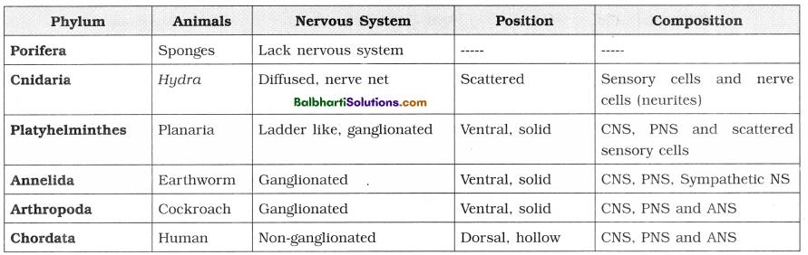
Nervous System in Hydra-
- Hydra → diffused nervous system in the form of nerve net.
- Two nerve nets in the mesoglea, one connected towards the epidermis and second towards the gastro-dermis.
- Sensory cells scattered in the body wall and tentacles, but no sense organs
- No sensory and motor nerves.
Nervous System in Planaria (flatworm)-
- Primitive animal with a central nervous system (CNS) located on the ventral side of body.
- Mass of cerebral or cephalic ganglion appearing like an inverted U-shaped brain.
- Ventral pair of nerves arising from ganglia. Interconnected to each other by transverse nerve or commissure in a ladder like manner.
- The peripheral nerve plexus arising laterally from VNC.
Neural tissue-
1. Two types of cells in neural tissue – the neurons and the neuroglia or glial cells.
2. Nerve is bundle of axons. Outside the CNS, it is called nerve while inside it is called tract.
3. Types of nerves : Sensory (with sensory * fibres), motor (with motor fibres) and mixed * type (with both sensory and motor fibres).
4. Neurons/Nerve cells :
- Neuron is structural and functional unit of * the nervous system.
- Each multipolar neuron has three parts – cyton : or cell body, dendron and axon.
5. Grey matter and white matter :
- Grey matter is darker part of CNS. This is due to presence of cytons.
- White matter is lighter part of CNS. This is due to presence of myelin sheaths around axons.
In PNS however, the accumulation of cyton causes a swelling on the nerve. Such a swelling is called ganglion, [cytons within CNS form nuclei while those in PNS form ganglia]
6. Connective tissue layers in a nerve are :
- Endoneurium : covers each nerve fibre
- Perineurium : covers each nerve bundle having a number of neurons
- Epineurium : covers many nerve bundles to form a peripheral nerve
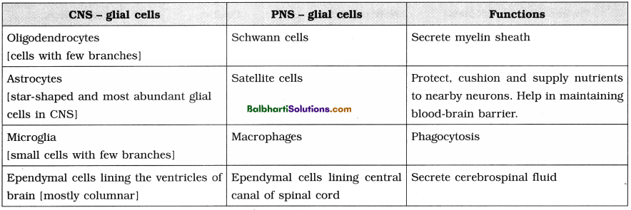
7. Neuroglial cells :
- More in number than the neurons.
- They are supporting cells of the Central Nervous System (CNS) and Peripheral Nervous System (PNS).
- Neurilemma is the plasma membrane of Schwann cell.
8. Tjrpes of neuroglial cells :
Synapse-
1. Junction between two nerve cells with a minute gap (synaptic cleft) in between them which allows transmission of impulse by a neurotransmitter bridge.
2. When the telodendria are connected to muscle fibre, it is called motor end plate or neuro¬muscular junction.
3. Properties of nerve fibres :
- Excitability/Irritability
- Conductivity
- Stimulus
- Summation effect
- All or none law
- Refractory period
- Synaptic delay
- Synaptic fatigue
- Velocity
4. Types of synapses : Electrical synapse and Chemical synapse.
(1) Electrical synapses are found in those places of the body requiring fastest response as in the defence reflexes.
(2) A chemical synapse between a motor neuron and a muscle cell is called a neuromuscular junction or motor end plate.
There are three components of a typical chemical synapse.
- The pre-synaptic terminal
- The synaptic cleft
- The post-synaptic neuron
5. Transmission of nerve impulse across a synapse :
- This transmission takes place with the help of neurotransmitters.
- Once the neurotransmitters bind to the receptors of the post-synaptic cell, the action is either excitatory or inhibitory depending on the type of neurotransmitter.
- The enzyme like acetyl cholinesterase destroys the neurotransmitter after the transmission and the synapse is ready to receive a new impulse.
Transmission of nerve impulse along the axon-
- The excitable neurons transmit the impulse through changes in electrical charges across the neuronal membrane.
- The external tissue fluid has both Na+ and K+ ions.
- This process is called sodium pump or Na-K exchange pump.
- Generation of nerve impulse : Occurs through depolarization.
- Saltatory conduction takes place in medullated nerve fibres.
![]()
Human Nervous System-
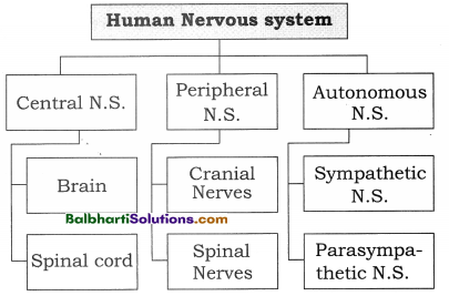
1. Central nervous system (CNS) :
- Brain is enclosed within the brain box/ cranium of the skull, whereas the spinal cord lies in the vertebral canal of the vertebral column.
- Inner to these bony structures, there are 3 protective membranes called meninges.
2. Meninges :
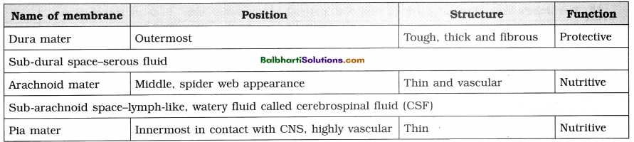
3. CSF (cerebrospinal fluid) :
- About 100-120 cc lymph like extra cellular . fluid with specific gravity of 1.005, present in and around the CNS.
- It is secreted by the pia mater, the choroid plexuses and the ependymal cells lining the ventricles of the brain and central canal of spinal cord.
4. Functions of meninges and CSF :
- Shock absorber, protection, prevention of desiccation.
- Maintaining constant pressure inside as well as outside the CNS.
- Exchange of nutrients and wastes between blood and brain tissue.
- Supply of oxygen to the brain.
5. The Human brain :
- Encephalology : Study of all aspects of the brain.
- About 1300-1400 g in weight -and 1300-1500 cc in volume
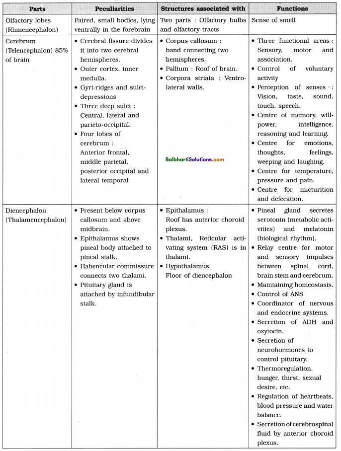
6. Functional areas of cerebrum :
| Areas | Functions |
| 1. Frontal lobe | Motor area → controls voluntary motor activities or movements of muscles. Premotor area → higher centre for involuntary movements and autonomus nervous system.Association area → coordination between sensation and movements. Broca’s area → motor speech area. |
| 2. Parietal lobes | Somaesthetic sensation of pain, pressure, temperature, taste |
| 3. Temporal lobes | Centres for smell (olfactory), hearing (auditory), speech and emotions. |
| 4. Occipital lobes | Visual area mainly for sense of vision. |
| 5. Wernicke’s area | Present partly in temporal, parietal and occipital lobes. Sensory speech area. |
| 6. Basal nuclei or basal ganglia | Control precise muscular activities at subconscious level. |
| 7. Corpus striatum | At the floor of cerebrum is the largest basal nucleus. |
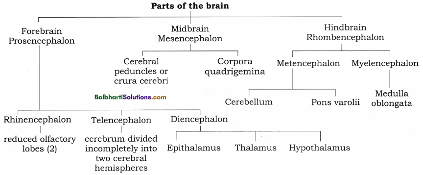
7. Parts of the Hindbrain (Rhombencephalon) :
8. Parts of the Midbrain (Mesencephalon):
9. Parts of the Hindbrain (Rhombencephalon) :
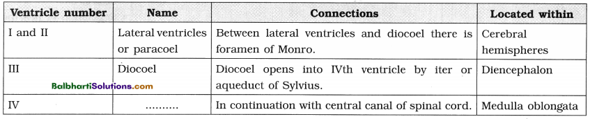
10. Ventricles of brain : The cavities present in the different parts of the brain are called ventricles.
![]()
11. Important terms associated with brain.
- Corpus callosum : Transverse band of nerve fibres which connects right and left cerebral hemisphere. It is the largest commissure of the brain.
- Cerebral cortex : The outer surface of cerebrum, composed of grey matter.
- Cerebral medulla : Inner part composed of white matter.
- Gyri (elevations) and Sulci (depressions) : convolutions and grooves on the surface of cerebrum.
- Central sulcus : Between frontal lobe and the parietal lobes.
- Parieto-occipital sulcus : Between parietal and occipital lobes.
- Lateral or Sylvian sulcus : Between temporal lobe and frontal and parietal lobes.
- Insula or insular cortex : Fifth lobe which is folded deep within the lateral sulcus.
- Foramen of Monroe : Narrow opening through which two lateral ventricles communicate with diocoel (third ventricle).
- Pineal gland : Vestigial 3rd eye and an important endocrine gland, producing hormones melatonin and serotonin.
- Habenular commissure : Connects two thalami.
- RAS (Reticular Activating System) : Relay centre as it transmits all sensory impulses except those of olfactory to the cerebrum. Situated in thalami.
- Aqueduct of Sylvius or iter : Connection between third and fourth ventricle through hypothalamus and midbrain.
- Limbic system : A complex neuronal circuit formed by the hypothalamus, amygdala, parts of epithalamus and thalamus, hippocampus and other areas.
- Optic chiasma : Crossing of the two optic nerves.
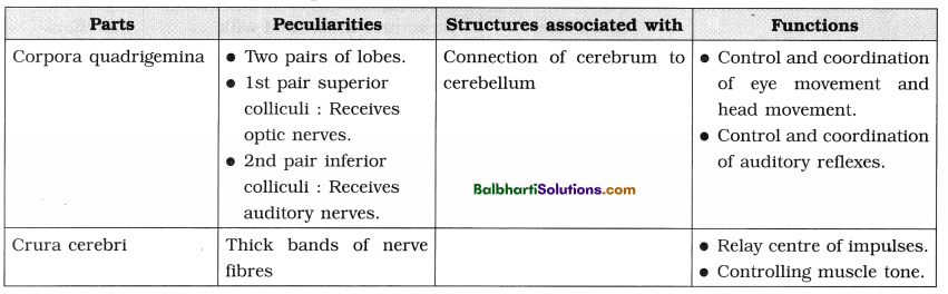
- Corpora quadrigemina : Four rounded elevations on the dorsal surface of the midbrain. The two superior colliculi are involved in visual reflexes and the two inferior colliculi are for auditory reflexes.
- Crura cerebri : Two thick fibrous tracks, also called cerebral peduncles, situated in the floor midbrain.
- Red nucleus : Grey matter near the centre of the midbrain, controlling posture. and muscle tone, modifying some motor activities and motor coordination.
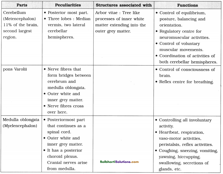
- Pons varolii : Rounded bulge on the underside of the brain stem.
- Brain stem : Consist of midbrain, pons and medulla.
- Arbor vitae : The mixing of white matter with the grey matter showing a branched tree-like pattern.
- Cerebellar peduncles : Three pairs of myelinated nerve bundles connecting cerebellum to the other parts of CNS.
- A pair of lateral – foramina of Luschka and a median – foramen of Magendie :
apertures on the posterior choroid plexus.
12. Spinal Cord :
- Spinal cord is the lower extension of the medulla oblongata of the brain.
- It lies within the neural canal of the vertebral column and is surrounded by three meninges.
- Externally, the spinal cord appears as long cylindrical rod.
- It is 42 to 45 cm long and 2.0 to 2.5 cm broad.
- Conus medullaris : Terminal nervous part of the spinal cord.
- Filum terminate : Thread like non-nervous extension.
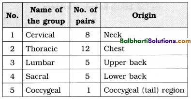
- 31 pairs of spinal nerves arise from lateral sides of the spinal cord.
- Cauda equina – Filum terminale with some spinal nerves running parallel to it. (appearing like a horse-tail)
13. T. S. of spinal cord :
- The spinal cord has a deep, narrow dorsal fissure and a broad ventral fissure.
- The inner grey matter is H-shaped and the outer white matter surrounds it.
- Grey matter is divisible into six horns, namely dorsal, lateral and ventral horns.
- The white matter is divisible into 6 columns or funiculi, namely dorsal, lateral and ventral funiculi.
- The dorsal and ventral horns extend out of the spinal cord as dorsal root and ventral root.
- The dorsal root has dorsal root ganglion which is a collection of unipolar sensory neurons. No such ganglia on ventral root.
- The adjustor/association or inter-neurons lie inside the grey matter.
- The white matter consists mainly of bundles of myelinated nerve fibre called ascending and descending tracts.
Functions :
- The spinal cord is the main centre for the most reflex actions.
- It provides pathway for conduction of sensory and motor impulses.
14. Peripheral Nervous System (PNS) : The peripheral nervous system connects the central nervous system to the different parts of the body having receptors and effectors.
Two types of peripheral nerves :
- Cranial nerves : arise from the brain.
- Spinal nerves : arise from the spinal cord.
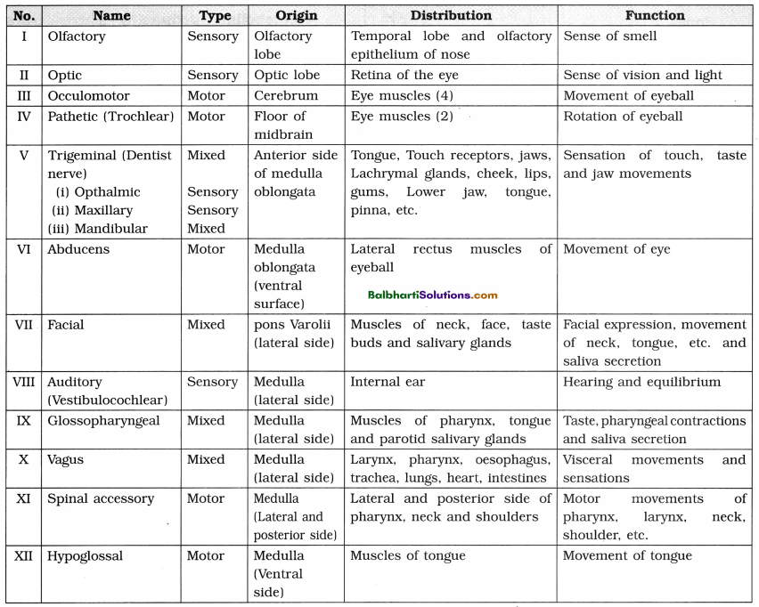
15. Cranial Nerves :
16. Spinal Nerves:
- Thirty-one pairs of spinal nerves originate from the spinal cord.
- Spinal Nerves : All spinal nerves are mixed nerves.
17. Formation of a typical spinal nerve :
- Each spinal nerve is formed inside the neural canal of vertebral column.
- The dorsal sensory and the ventral motor nerves together form the mixed spinal nerve.
- As soon as it emerges out of vertebral column, it shows three branches, viz.
a. Ramus dorsalis
b. Ramus ventralis
c. Ramus communicans
18. Reflex Action :
(1) It is a quick, automatic, involuntary and spontaneous response to stimulus,
(2) The path along which the action is carried out is called reflex arc.
(3) Components of a reflex arc :
a. Receptor/sense organ
b. Sensory/afferent neuron
c. Association/adjustor neuron
d. Motor/efferent neuron
e. Effector organ
(4) Types of reflexes :
a. Somatic and visceral
b. Cranial and spinal
c. Simple [monosynaptic] and complex [polysynaptic]
d. Unconditional and conditional
table
(5) According to recent studies, the ANS is under the control of CNS and nerves arising from it (PNS).
According to this view, the PNS is divided into
(i) Somatic nervous system
(ii) Autonomic nervous system
19. Autonomic Nervous System (ANS) :
- Autonomic nervous system transmits impulses from CNS to the involuntary organs and smooth muscles of the body.
- It includes – autonomic ganglia,
preganglionic fibres and postganglionic fibres. - Autonomic ganglia include
- Sympathetic ganglia – present near CNS in the form of sympathetic cord.
- Parasympathetic ganglia – present near or on the effector organs.
- Preganglionic fibres arise from grey matter of CNS and end at autonomic ganglia.
- Postganglionic fibres arise from autonomic ganglia to the effector organs.
Autonomic nervous system consists of sympathetic and parasympathetic nervous system.
(1) Sympathetic Nervous System (SNS) :
- Also called thoraco-lumbar outflow.
- Consists of 22 pairs of sympathetic ganglia which lie near vertebral column.
- Post ganglion is neuron which produce adrenaline. Hence they are called adrenergic fibres.
- It works in emergencies. It is also called 3 Fs system [fright, fight and flight]. It has excitatory and stimulating effect on most organs of the body.
![]()
(2) Parasympathetic Nervous System:
- It is also called cranio-sacral outflow.
- It consists of ganglia which are very close or within the wall of the effector organs.
- Acetyicholine is produced at the terminal end of postganglionlc nerve at the effector organ. Hence these are also called cholinergic fibres.
- All activities which are stimulated by the sympathetic system are brought back to normal by this system. Hence it is also called housekeeping system.
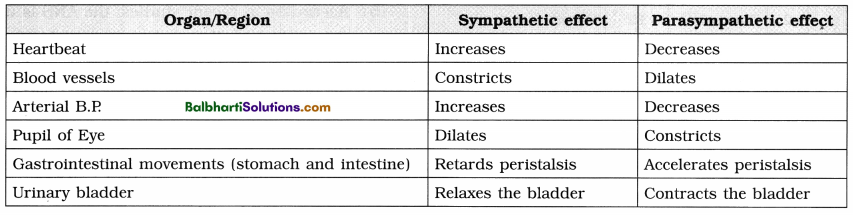
Comparison between Sympathetic and Parasympathetic Nervous System :
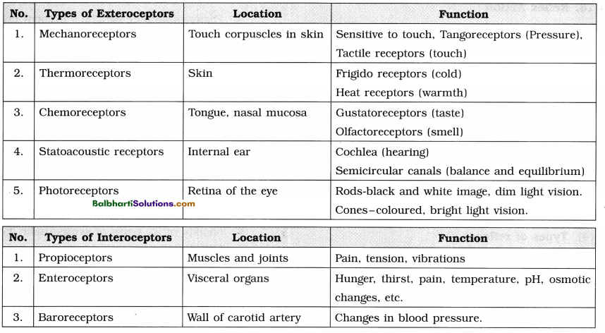
Sensory Receptors-
1. Specialised structures in the body modified to receive the various stimuli from the external or internal environment.
2. Classification of receptors : Receptors are classified on the basis of their location, function and their sensitivity to specific stimuli. Their classification is given in the following chart.
Types of exteroceptors and interoceptors, their locations and functions :
3. Eye :
- The eyes are a pair of sensory organs of vision located in the orbit of skull.
- Each eye is spherical/rounded and called eyeball.
- Wall of the eyeball is made up of 3 layers : (1) sclera, (2) choroid (3) retina.
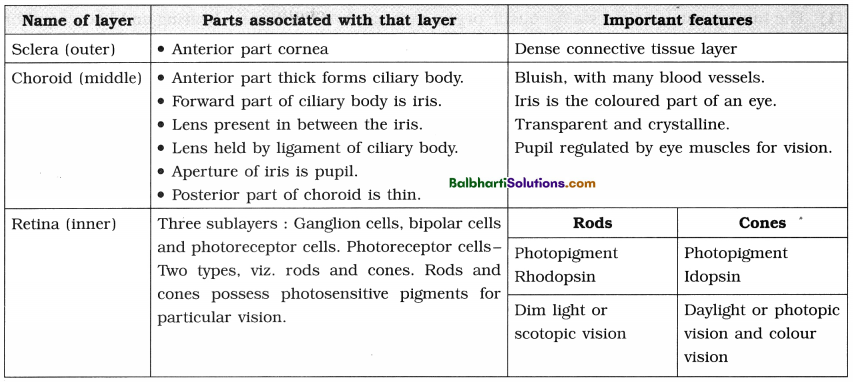
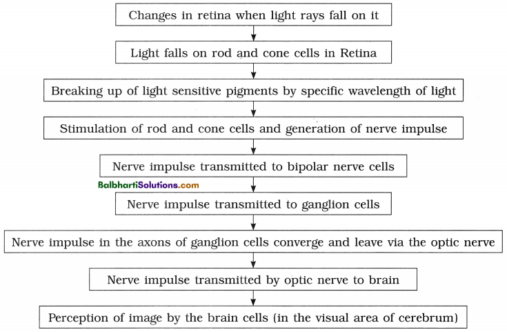
Generation of image/Mechanism of vision :
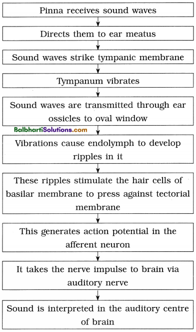
4. Ear :
- The human ear is called statoacoustic organ as it has two functions – hearing and body equilibrium.
- Anatomically the ear is made up of three divisions : the external ear, middle ear and inner ear.
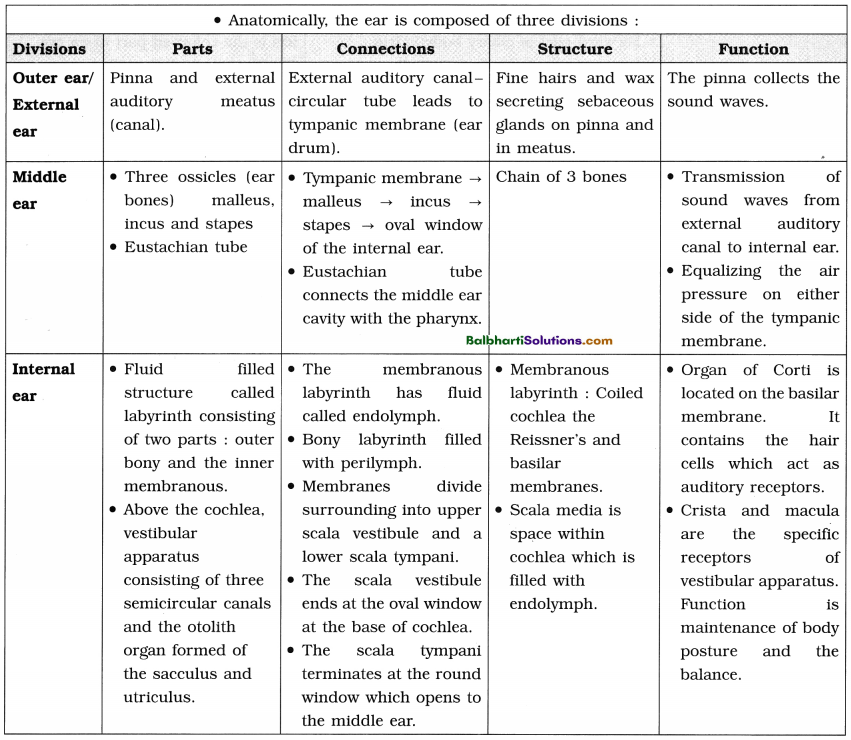
(3) The organ of hearing :
- Organ of Corti is a pea sized structure located on basilar membrane. It has a sensory epithelium over the basilar membrane.
- The sensory cells have sensory hair on their free end so also called hair cell. In between the rows of hair cells are present supporting cells.
- Hair cells have long stiff microvilli called stereocillia on their apical surfaces. Above these stereocilia, is a jelly like membrane called tectorial membrane.
- This organ acts as a transducer, converting sound vibrations into nerve impulses.
![]()
(4) Other parts of the ear :
- Besides the cochlea, the internal ear also has the vestibular apparatus.
- It is composed of three semi-circular canals and the utriculo saccular region.
- All three semi-circular canals lie in different planes at right angles to each other.
- These canals are filled with endolymph. The base of each of the canal has an ampulla in which there is a sensory ridge called crista. The structure is crista ampullaris.
- The vestibule has two sensory spots – macula of saccule and utricle. The utricle is larger than saccule.
(5) Mechanism of Hearing :
Disorders of nervous system-
1. Psychological disorders :
Commonly called mental disorders. There is a wide range of conditions that affect the mood, thinking or behaviour.
Some of the major categories of psychological disorders are :
- Intellectual disability (earlier known as mental retardation),
- Autism spectrum disorder
- Bipolar disorder
- Depression
- Anxiety disorder
- ADHD (Attention Deficit Hyperactivity Disorder)
- Stress related disorders.
2. Parkinson’s disease :
- Degeneration of dopamine-producing neurons in the CNS causes Parkinson’s disease.
- Symptoms develop gradually over the years.
- Symptoms are tremors, stiffness, difficulty in walking, balance and coordination.
3. Alzheimer’s disease :
- It is the most common form of dementia.
- Its incidence increases with the age.
- Symptoms include the loss of cognitive functioning- thinking, remembering, reasoning and behavioural abilities. It interferes with the person’s daily life and activities.
Chemical Coordination
Endocrine system –
1. The cells in organisms communicate with each other through chemical signals. These cells are broadly of four types as follows :
- Autocrines : Cells release secretion to stimulate themselves.
- Paracrines : Cells release secretion to stimulate neighbouring cells.
- Endocrines : Cells release secretion to stimulate distant cells.
- Pheromones : Cells/Organs release secretions to stimulate other organism.
2. Chemical coordination is carried out by secretions of ductless glands or endocrine glands. Hence this chemical coordination system is also called the endocrine system.
3. Endocrine system :
- The endocrine system controls body activities by means of chemical messengers called hormones.
- Hormones are released directly into the blood.
4. Properties of Hormones :
- They act as chemical messengers and are effective in very low concentration.
- Hormones can function as regulators that inhibit or stimulate or modify specific processes.
- Hypersecretion or Hyposecretion of hormones leads to various disorders.
- These are metabolised after their function and are excreted through urine.
5. Mechanism of hormone action :
- Hormones are released in a very small quantity.
- They produce their effect on the target organs/cells by binding to hormone receptors.
- The hormone receptors may be on the cell membrane or may be intracellular.
- A hormone receptor complex is formed and . this leads to biochemical change in the target tissue.
(a) Mode of hormone action through membrane receptors :
- Hormones like catecholamines, peptide and polypeptide hormones are not lipid soluble. Therefore they cannot enter their target cells through plasma membrane.
- Molecules of amino acid derivatives, peptide hormones bind to specific receptor molecules located on the plasma membrane.
- The hormone receptor complex causes the release of an enzyme adenylate cyclase from the receptor site. This enzyme forms cyclic AMP from ATP of the cell.
- The hormone acts as the first messenger and cAMP is the second messenger.
(b) Mode of hormone action through intracellular receptors :
- Steroid and thyroid hormones are lipid soluble and easily pass through plasma membrane of target cell into the cytoplasm.
- In the cytoplasm, they bind to specific intra¬cellular receptor proteins forming a hormone-receptor complex that enters the nucleus.
- In the nucleus, the hormone receptor complex binds to a specific regulatory site of DNA.
Major endocrine glands-
1. Hypothalamus :
- Ectodermal in origin.
- Forms the floor of diencephalon.
- Major function is to maintain homeostasis.
- Controls the secretory activity of pituitary gland by the release and inhibiting hormones.
- All hormones of hypothalamus are peptide hormones.
2. Hormones of hypothalamus :
- Somatotropin/GHRF
- Somatostatin/GHRIF
- Adrenocorticotropin Releasing Hormone
- Thyrotropin Releasing Factor .
- Gonadotropin Releasing Hormone (GnRH)
- Prolactin Inhibiting Hormone (Prolactostatin)
- Gastrin Releasing Peptide (GRP)
- Gastric Inhibitory Polypeptide (GIP)
3. Pituitary gland or hypophysis :
(1) External morphology :
- Pea sized reddish-grey coloured gland.
- Controls almost all other endocrine glands, hence previously it was called the master endocrine gland.
- However, hypothalamus controls it through the releasing and inhibiting factors.
- Located just below the hypothalamus and is attached to it by a stalk called infundibulum or hypophyseal stalk.
- Remains lodged in a bony depression called sella turcica of the sphenoid bone.
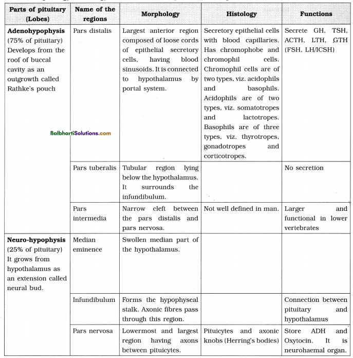
- Consists of two lobes called anterior lobe (Adenohypophysis) and posterior lobe (Neurohypophysis). Intermediate lobe (Pars intermedia) is a small reduced part lying in the cleft between the anterior and posterior lobe.
- Neurohypophysis is connected directly to the hypothalamus by axon fibres forming hypothalamo- hypophyseal tract,
- Adenohypophysis and intermediate lobe are connected to the hypothalamus through hypothalamo- hypophyseal portal system.
(2) Parts, morphology, histology and functions of pituitary in a glance :
![]()
(3) Hypothalamo – Hypophyseal portal system :
- Various hormones secreted by hypothalamus reach the pituitary gland through this portal system.
- The portal vein collects blood from various parts of hypothalamus and opens into anterior lobe of pituitary.
- From pituitary, the vein finally carries the blood into the superior vena cava.
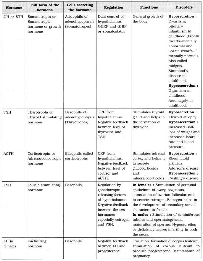
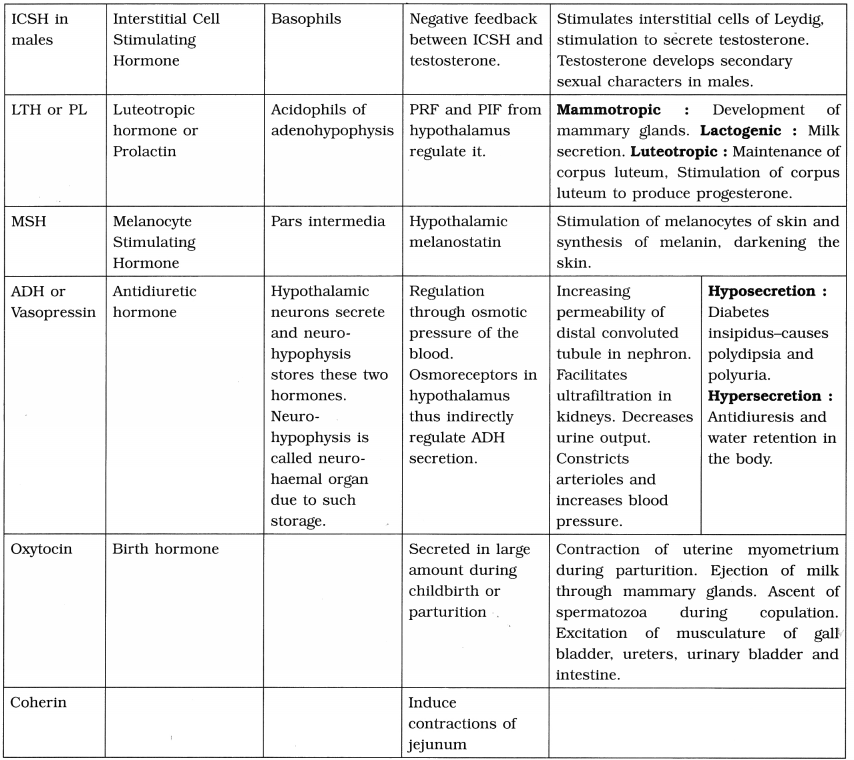
(4) Hormones of pituitary and their role :
4. Pineal gland :
- The pineal body/pineal gland is given off from the roof of diencephalon. It is located between the two cerebral hemispheres.
- The pineal gland is sensitive to the biochemical signals of light.
- It secretes a hormone called melatonin also known as sleep hormone.
5. Thyroid gland :
(1) Morphology :
- It is the largest endocrine gland.
- The two lobes of thyroid gland are connected a non-secretory band called isthmus.
(2) Internal structure :
- The thyroid lobes are composed of rounded follicles held together by interfollicular connective tissue called stroma.
- The stroma contains blood capillaries and small group of parafollicular cell or ‘C’ cells.
- Thyroid follicles Eire composed of cuboidal epithelium resting on a basement membrane and is filled with a gelatinous colloid.
(3) Thyroid hormones :
- The two hormones secreted by the follicular cells are Thyroxine/tetra iodothyronine/T4 (four atoms of iodine) and Triiodothyronine or T3 (three atoms of iodine).
- Parafollicular cells produce a hormone thyrocalcitonin whose production is not under the control of TSH.
(4) Formation of T3 and T4 :
- Thyroxine is synthesized by attaching iodine to amino acid tyrosine by enzymatic action.
- The amino acid tyrosine molecule binds to iodine to produce Monoiodotyronine (T1) or 2 atoms of iodine to produce Diiodothyroninc (T2).
- T1 and T2 molecules bind end to end to make colloidal mass inside the follicle. They are further metabolised to prepare T3 and T4.
(5) Functions of Thyroid hormones :
- Regulation of the basal metabolic rate of body.
- Regulation of metabolism by stimulating protein synthesis and promotes growth of body tissues.
- Calorigenic effect as it helps in thermoregulation by increasing heat production.
- Increases action of neuro transmitters – adrenaline and nor adrenaline.
- Supports the process of RBC production and maintenance of water and electrolyte balance.
- Regulates reproductive cycles in females.
- Parafollicular cells or ‘C’ cells produce thyrocalcitonin hormone, which regulates calcium metabolism.
- Calcitonin is the active form of hormone, which is hypocalcemic hormone. It regulates the concentration of calcium and phosphorus in the blood.
![]()
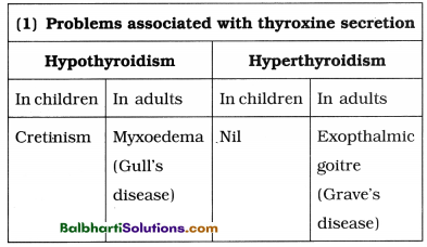
(6) Disorders related to thyroid gland :
6. Parathyroid gland :
- Situated on the posterior surface of the lobes of thyroid gland.
- 2 pairs, named as superior and inferior parathyroid glands.
- Cells are arranged in a compact mass.
(4) Hormones :
- The parathyroid secretes a peptide hormone called parathormone (PTH). It is also called
Collip’s hormone. - Regulates calcium and phosphate balance between blood and other tissues. It is a hyper calcemic hormone. Release of parathormone increases blood calcium level.
- It stimulates osteoclast of bones to stimulate bone resorption.
- Thus, parathormone and calcitonin are antagonistic hormones.
(5) Disorders :
| Hyposecretion of parathormone | Hypersecretion of parathormone |
| Parathyroid tetany or hypocalcaemic tetany. | Osteoporosis. |
| Lowers concentration of calcium in the blood. This increases excitability of nerves and muscles causing muscle twitch and spasm. | Responsible for more resorption of calcium from bones i.e., demineralization of bones resulting in softening, bending and fracture of bone. |
7. Thymus gland :
(1) Located in the upper part of thorax on the dorsal side of the heart just behind sternum.
(2) Prominent gland at birth till puberty but gets gradually atrophied in the adult due to action of sex hormones.
(3) Functions:
- Secretes the hormone thymosin.
- Important role in the development of immune system by maturation of T-lymphocytes.
8. Adrenal gland/Suprarenal gland:
(1) Adrenal glands have dual origin from mesoderm and ectoderm.”
(2) Located on the upper border of each kidney.
(3) Small. conical yellowish glands having two distinct regions, outer cortex and inner medulla.
| (A) Adrenal cortex (outer) | (B) Adrenal medulla (inner) |
| Derived from embryonic mesoderm. Secretes many hormones together called corticoids. Main two hormones : (1) Glucocorticoids (2) Mineralocorticoids | Derived from embryonic ectoderm. Secretes main two hormones (1) Adrenaline (epinephrine) (Emergency hormone, also called 3F hormone – (fight, flight and fright). (2) Noradrenaline (norepinephrine). (Regulates the blood pressure under normal condition, acts as vasoconstrictor) |
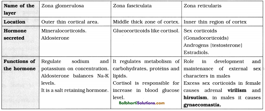
(4) Three concentric regions of adrenal cortex :
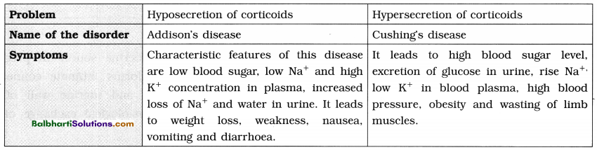
(5) Disorders related to Adrenal cortex :
9. Pancreas :
- Develops from endoderm.
- It is heterocrine i,e. both exocrine and endocrine gland.
- Endocrine cells of pancreas form groups of cells called Islets of Langerhans.
- There are four kinds of cells in islets of
Langerhans which secrete hormones.
- Alpha (α) cells (20%) secrete glucagon.
- Beta (β) cells (70%) secrete insulin.
- Delta ((δ) cell (5%) secrete somatostatin
- PP cells or F cells (5%) secrete pancreatic polypeptide (PP).
Disorder related to pancreas : Diabetes mellitus
- Hyperglycemia i.e. It leads to increased blood glucose level.
- Cause : Under activity of Beta cells, which results in reduced secretion of insulin.
- Types of diabetes :
(1) TYPE-1 diabetes i.e. insulin dependent diabetes mellitus (IDDM)
(2) TYPE-2 diabetes i.e. Non insulin dependent diabetes mellitus (NIDDMj.
- Diabetes causes glucosuria, excessive urination and dehydration of body tissues, degradation of fats and increase in formation of ketone bodies (ketosis).
- Administration of insulin lowers blood glucose level.
10. Gonads : Gonads are sex organs (the testes and the ovaries).
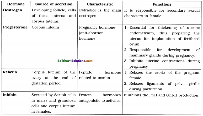
(1) Ovaries :
(2) Testes : Testes secrete male sex hormones called androgens such as testosterone.
Testosterone :
- It is secreted from interstitial cells or Leydig cells by the influence of luteinising hormone (LH).
- Rise in testosterone level in blood above normal inhibits LH secretion.
- It is also responsible for appearance of secondary sexual characters such as facial and pubic hair, deepening of voice, broadening of shoulders, male aggressiveness, etc.
![]()
11. Placenta :
- Temporary endocrine source in pregnant women which forms intimate connection between foetus and uterine wall of the mother for physiological exchange of the materials.
- During pregnancy, placenta secretes hormones such as estrogen, progesterone, HCG (Human Chorionic Gonadotropin) and human placental progesterone.
12. Diffused endocrine glands :
(1) Gastro-intestinal tract : Certain cells of gastrointestinal mucosa are endocrine in function. Their hormones play vital role in digestive processes and flow of digestive juices.
| Hormone of GI tract | Function |
| 1. Gastrin | Stimulates gastric glands to produce gastric juice. |
| 2. Secretin | Responsible for secretion of pancreatic juice and bile from pancreas and liver. |
| 3. Cholecystokinin CCK/ Pancreozymin PZ : | Stimulates gall bladder to release bile and stimulates the pancreas to release its enzymes. |
| 4. Entero-gastrone/Gastric inhibitory peptide (GIP) | Slows gastric contractions and inhibits the secretion of gastric juice. |
(2) Kidney :
- Hormones of kidney – renin, erythropoietin and calcitriol (calcitriol is the active form of vitamin cholecalciferol -D3).
- Renin along with angiotensin helps in maintaining the blood pressure in the renal artery by vasoconstriction.
- Erythropoietin stimulates erythropoiesis.
- Calcitriol helps in absorbing calcium from the stomach.
(3) Heart :
- Hormone of heart-Atrial natriuretic Factor /ANE
- Increases sodium excretion [natriuresis] along with water by kidneys.
- Reduces blood pressure by lowering blood volume.
13. Hormone therapy/HT :
- Use of hormones in medical treatment.
- Required for the patients during pregnancy, menopause, osteoporosis, growth hormone deficiency, insulin resistance, cancer, etc.